It's no secret that CT radiation dose levels in children -- especially in the emergency department, where patients are fidgety and physicians need fast answers -- are too high.
A recent observational study pegged the CT radiation dose range at 6 to 10 mSv, according to Dr. Patrik Rogalla, professor of radiology at the University of Toronto. Even more concerning is the wide range cited in the study: 0.7 to 26 mSv, he said in a presentation at the 2009 RSNA meeting in Chicago.
"What we're seeing is because ED physicians don't want sedation, they are scanning several times until they get one right," Rogalla said. "They didn't know the patient was moving and they [rescanned] again and again."
One way to stop motion at low-dose CT is to speed up the scan. Several presentations at the 2009 RSNA meeting used dual-source CT and high-pitch scanning to minimize motion artifacts in pediatric imaging, for example.
Another solution -- the one chosen by radiologists at the University of Toronto and Charité Medical University in Berlin, who performed the present study -- is the use of wide-area-detector scans with 320-detector-row CT. Each rotation yields 16 cm of anatomic coverage, enough to stop the fidgeting and cover the target in most patients in a single rotation.
To assess the scanner's dose and image quality, they examined scans performed on a total of 39 young patients with abdominal complaints (mean age, 2.1 years; median, 0.8 years; range, 1 day to 14 years). All were scanned with dynamic volume CT on a 320-detector-row scanner (Aquilion One, Toshiba America Medical Systems, Tustin, CA).
Settings for the study included 80 kV for contrast-enhanced scans, 120 kV for noncontrast scans, and 10-50 mA and 0.35-0.5 sec gantry rotation time, with one to three rotations depending on the target.
Milliamperes were calculated using the following formula: (body weight [in kg] + 5) x f, with f = 1 for chest and f = 1.5 for abdominal scans at 120 kV. For 80-kV scans, radiologists multiplied the mAs value by a factor of 2.5.
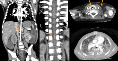
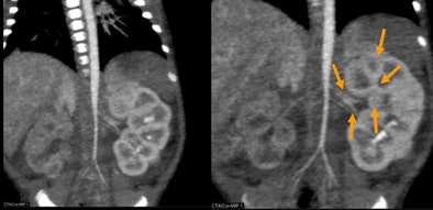 |
| Newborn with aortic hypoplasia and renal artery stenosis. Weight of 1.7 kg; single-rotation 320-detector-row scan at 0.5 sec, 80 kV, 40 mA, and dose of 0.3 mSv. All images courtesy of Dr. Patrik Rogalla. |
"We varied mAs by patient weight, and sometimes we [needed] one or two rotations depending on the patient," Rogalla said. For noncooperative patients and those who were unable to hold their breath, as many as three rotations at 0.35 seconds were acquired to shift the reconstruction within the acquisition window for motion artifact reduction. Every study used the 640-detector-row reconstruction mode (0.5 mm) available on the scanner.
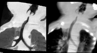 |
| Patient with stridor and giant cervical hemangioma was breathing continuously and not sedated during 320-detector-row CT, acquired in two rotations at 0.35 sec, 80 kV, 20 mA, and dose of 0.3 mSv. Below, additional rotation allows reconstruction of images at multiple time points, revealing air trapping that would have been missed at single-rotation scan. |
 |
Two expert readers evaluated all scans with respect to image quality on a scale of 1 to 3 (1 = poor). Radiation dose was calculated based on the dose-length product (DLP) displayed on the dose report and verified using commercially available software (CT-Expo, version 1.51d), Rogalla said.
In all but four patients (two chest and two abdominal scans), the 16-cm detector coverage sufficed for scanning the target area in a single gantry rotation. No scan was rated poor, and one patient moved despite manual fixation, so a repeat scan was needed, the authors wrote in an abstract.
Despite patients' continuous breathing during image acquisition, axial slices were rated good. In one patient, motion blur was relevant but did not prevent a diagnosis. Radiation exposure (calculated by both the readout and sensors) ranged from 0.2 to 2.3 mSv (mean, 0.6 mSv), depending on the scanning area and parameters used.
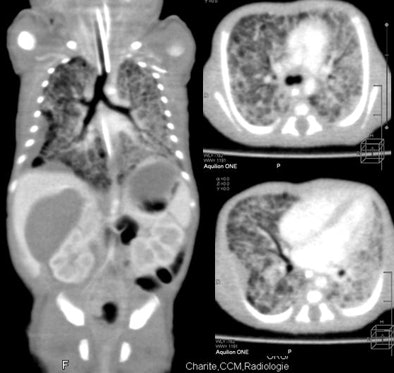 |
| Nearly whole-body imaging in a single rotation reveals lung changes and pleural thickening. |
There were several limitations to the study, including a lack of comparison to helical scans and a very young cohort. "We did have predominance of neonates, so we have to be cautious in generalizing exposures rates," Rogalla said of the results.
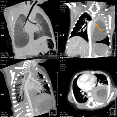 |
| Feeding artery is visible at 320-detector-row CT in a patient with pulmonary blastoma. |
"A dynamic volume 320-slice CT ... potentially may reduce the overall radiation dose if used correctly," he said. "In particular, we have reduced motion artifacts with ultrafast flash imaging -- so even though the patients were breathing and moving, we didn't have significant motion artifacts because it's so fast."
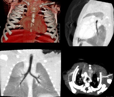 |
| Vascular ring and hypothesis of tracheal stenosis. Images at 320-detector-row CT. |
Fast scanning reduces the need for sedation, which contributes to overall reduced risk for the patient, he added.
An audience member asked if this kind of scanning protocol could realistically be expected to be a dominant force in future imaging.
"My opinion is that the trend is toward wide-area CT," Rogalla said. "So one rotation, half rotation, flat imaging is the way to go. I think there's great potential for reducing the dose."
By Eric Barnes
AuntMinnie.com staff writer
March 3, 2010
Related Reading
Study finds wide variations in global pediatric CT dose, February 25, 2010
CATCH rules accurately predict which kids need head CT, February 9, 2010
320-row perfusion CT shows promise for pancreatic tumors, February 5, 2010
Christmas ornaments can be a holiday hazard for toddlers, December 22, 2009
California technologist faces testimony in CT overdose case, September 18, 2009
Copyright © 2010 AuntMinnie.com


