Dear Cardiac Imaging Insider,
The use of CT perfusion (CTP) to assess myocardial viability is still a pretty new technique, and as such, comes with a few technical limitations. But its promise of gauging the functional significance of coronary artery stenosis without a second exam has gotten the attention of researchers in Munich.
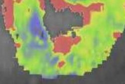
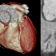
Putting CTP to the test in symptomatic patients who also underwent cardiac MRI, the team from Grosshadern, Germany, found that CT did a good job of detecting myocardial hypoperfusion -- and an even better job of finding myocardial infarction. What do researchers think will be needed to perfect the technique? Find out this and more in today's cardiac imaging story.
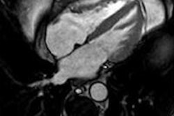
In other news from the heart, diastolic dysfunction is even a little more common than researchers thought -- and more prevalent among men than women, concluded a research team from Portugal that looked at MRI results in patients with suspected heart disease. Because subclinical diastolic dysfunction can increase the risk of heart failure, it's definitely worth knowing about as early as possible, the investigators said. Get the rest of the story here.
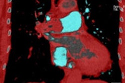
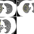
Pooled-blood volume (PBV) dual-energy CT (DECT) can be a powerful tool for separating clinically important pulmonary embolism (PE) from less important filling defects that won't impact the patient, explained researchers from St. George's Hospital in London. And while most multidetector-row CT scanners can evaluate filling defects, DECT goes a step further by suggesting the appropriate treatment for the defect at hand. Most physiologically impairing occlusive PE are associated with perfusion defects on PBV imaging, while nonimpairing pulmonary hypertension cases do not cause perfusion defects at PBV imaging, the group wrote in an article you'll find here.
On the other hand, dual-source CT lends no particular advantage over single-source in transaortic valve implantation (TAVI) planning procedures, according to another study from the U.K. And importantly for clinicians, the double scan previously required -- one of the heart and a second scan of the thorax to include the access vessel for TAVI/TAVR -- has been rolled into a single exam that can be acquired on any advanced scanner, the German study team said.
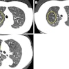
In recent years, doctors have grown increasingly concerned about the effects of radiotherapy for cancer on cardiac function. Is it time to scan the hearts of patients who have been treated with radiotherapy for diseases such as lung cancer? A Belgian study team makes the case for imaging in an article you'll find here.
Dutch researchers are finding that not all chest pain patients need coronary CT angiography. Learn when it's safe to say no, right here.
Finally, we invite you to scroll down through the links below for more news about cardiac imaging in Europe, all in your Cardiac Imaging Digital Community.


