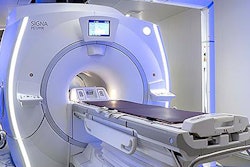
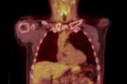
VIENNA - FDG-PET/MRI can produce diagnostic quality images "comparable" to FDG-PET/CT in patients with head and neck cancer, according to a study presented on Thursday during the opening day of ECR 2013.
Researchers from the University of Leipzig in Germany found no significant statistical difference regarding sensitivity, specificity, and positive and negative predictive values between the two hybrid modalities.
The study, led by Dr. Patrick Stumpp from the department of diagnostic and interventional radiology, evaluated 17 patients (13 males and four females) with a mean age of 60 years, ranging from 42 to 78 years. Seven patients were sent for primary staging and 10 patients for restaging.
Patients first received an FDG-PET/CT scan 90 minutes after injection, with image acquisition at three minutes per bed position and imaging done from the skull to the thigh. The FDG-PET/MRI (Biograph mMR, Siemens Healthcare) exam began approximately 20 minutes later, with imaging at five minutes per bed position from the vertex to the thigh.
"When we were done with that, patients got a dedicated MRI scan of the head and neck region, which takes approximately 25 minutes with all the [imaging] sequences," Stumpp explained during his presentation. "For the first 10 minutes, we also gathered information for the PET [image] as well."
Image assessment
The PET images were independently evaluated by two physicians, while two radiologists interpreted the CT and MR images. All readers were blinded to any clinical information. Two teams of one radiologist and one nuclear medicine physician each reviewed the combined FDG-PET/CT and FDG-PET/MRI results.
"The people on these teams were instructed to search for the tumor tissue, either as the primary tumor, recurrence, lymph nodes, or metastases," Stumpp said.
The analysis was compared with histology results, which were available in nine cases. In the remaining eight subjects, consensus reading was used when all clinical and head and neck surgical information was available.
Stumpp and colleagues found 23 lesions, eight (35%) of which were primary lesions and 15 (65%) of which were metastases. Also, 55 benign findings were determined to be inflammatory or nonspecific in histology or follow-up.
In comparing sensitivity, specificity, positive predictive value, and negative predictive value results between the two reader groups, the researchers found no statistical difference in their evaluations, according to Stumpp.
| Reader group 1 | Reader group 2 | |
| PET/CT | ||
| Sensitivity | 78% | 87% |
| Specificity | 85% | 89% |
| Positive predictive value | 71% | 75% |
| Negative predictive value | 91% | 94% |
| PET/MRI | ||
| Sensitivity | 78% | 83% |
| Specificity | 82% | 94% |
| Positive predictive value | 65% | 86% |
| Negative predictive value | 91% | 94% |
The PET image alone from the PET/CT scan achieved the greatest sensitivity of 96%, while the separate MR image from the PET/MRI exam resulted in the greatest specificity of 96%.
Based on the small sample of 17 patients, Stumpp and colleagues concluded that FDG-PET/MRI is "comparable" to FDG-PET/CT for detecting head and neck cancer, but larger studies should be conducted to confirm the results.
"We might see some advantage of PET/MRI among some [patient] subgroups. Personally, I think, especially with tumor recurrence, PET/MRI might help more than PET/CT," Stumpp said. "Before we had the PET/MRI scanner, we always hoped that MRI would improve that even more, but the [results] show that PET/CT is already very good, and there are probably some patients who have some advantage with an examination by PET/MRI."


