Better and faster imaging enables more accurate diagnoses with less risk and at lower cost than ever before. Faster scanning techniques also mean a change from static to dynamic information, while increasing computer power and faster and more complex postprocessing algorithms can deal with enormous datasets and provide new types of information, such as functional imaging.
"These changes require new expertise from radiologists," said Dr. Erik Ranschaert, a radiologist at the Jeroen Bosch Ziekenhuis (JBZ), 's-Hertogenbosch, the Netherlands, and a speaker at this afternoon's refresher course on image sharing. "We need to be aware of the entire disease process and able to analyze this dynamic and functional information. Multidisciplinary collaboration is also a prerequisite for useful integration of these new imaging techniques with the patient's treatment. For example, cardiologists are increasingly using noninvasive imaging techniques, and advanced postprocessing with supercomputer technology will eventually allow them to choose less invasive procedures. These are exciting developments."
An increasing number of surgical interventions use computer-based image-guided navigation techniques, such as computer-assisted orthopedic and spinal surgery (see www.caos-international.org), computer-assisted head and neck and ear, nose and throat surgery, image-guided neurosurgery, and minimally invasive cardiovascular and thoracoabdominal surgery. Also, the increasing development of surgical laparoscopy pushes for a better organization of image distribution because it is of major importance for preoperative planning and intraoperative guidance, he noted.
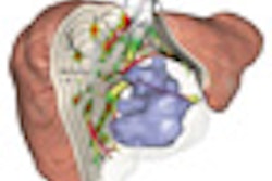
In oncology, there is increasing integration of imaging, particularly interventional radiology, with treatment in several areas: CT/MR diffusion and perfusion for planning and follow-up of tumor treatment -- e.g., to differentiate between necrotic and viable parts of the tumor; 3D segmentation for planning of treatment of liver tumors; robotics used for automated navigation of a needle or other instrument in brain surgery, surgery, and radiation oncology; interventional techniques for embolization of tumors, TACE (transarterial chemoembolization), radioembolization, intra-arterial chemotherapy, radiofrequency ablation (RFA), cryoablation, etc.
This trend has contributed to the formation of multidisciplinary societies such as the Society of Cardiovascular Computed Tomography (SCCT), the International Cancer Imaging Society (ICIS), the Intraoperative Imaging Society (IOIS), the Society for Molecular Imaging (SMI), and others.
At the JBZ, the new operating suite is being equipped with large screens that can be pulled down from the ceiling so that surgeons can make better use of imaging, as the diagram shows. Real-time guidance using ultrasound also will be offered. Surgery is becoming more and more microinvasive due to the use of endoscopic techniques, and precise guidance is increasingly important. Radiologists and surgeons will work together closely in areas such as selective tumor ablation using RFA with ultrasound, followed by partial liver resection.
Sharing images with colleagues outside of a radiologist's own hospital is also becoming easier, according to Ranschaert. For most second opinions offered in secondary or tertiary centers, previous imaging studies are still transferred on CD or DVD, but a more efficient and less costly solution is to make images automatically available by storing them in a "virtual cloud" that is accessible to other healthcare providers and patients. Several companies are already providing this type of service, especially in the U.S.
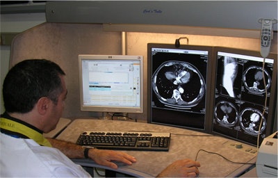
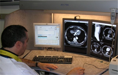 Reporting workstations at the University of Pisa allow multimodality image viewing and advanced image processing. Image courtesy of Dr. Davide Caramella.
More surgical and clinical/oncologic specialists will use their own software with navigation and visualization tools. Radiologists need to become more subspecialty oriented in order to be able to create added value in this process, and they need to focus on competence in specific areas, he said. They will also need to become more clinically oriented so that they are able to give useful advice. Treatment of the patient is increasingly complex and requires more intensive collaboration from all players involved. To be part of this imaging chain, from image acquisition to the report, they have to specialize and participate with other disciplines.
"We need to think more broadly and creatively," Ranschaert noted. "We should not limit our activities to just reading images from behind our workstation. Communication skills and a thorough knowledge of new treatment strategies are necessary. To increase awareness, it is important to incorporate presentations about this theme into meetings like the ECR."
An additional consideration for the future is imaging biomarkers, which raise the prospect of earlier detection of some diseases and promises to revolutionize basic research, drug development, and treatment. He observes that biomarkers are already enabling researchers to see in detail how candidate drugs are behaving, from determining the percentage of receptors occupied by a drug on target cells to looking at a drug's ability to cross the blood/brain barrier. This in turn can save time and money at the drug development lab bench. In particular, he is looking forward to new developments in target-specific MR contrast agents, which will allow the in vivo visualization of disease processes. MR contrast agents are being developed that are bound to specific targets, of which the distribution can be evaluated using MRI (e.g., liposomes with cholesterol and PEG-containing lipids).
At the University of Pisa in Italy, PACS was introduced 20 years ago, and for many years these systems served as "production tools'" designed to enhance the provision of radiological services. Soft-copy reporting and online access to images and reports from the clinical led to a reduction in turnaround time and helped physicians to make better use of imaging, explained session moderator Dr. Davide Caramella, from the department of radiology, Santa Chiara Hospital, Pisa. Only in recent years have radiologists and clinicians worked together in a real multidisciplinary environment.
At today's course, Caramella's surgical colleague, Dr. Andrea Pietrabissa, explained in detail about how the advent of minimally invasive surgery has made preoperative imaging assessment of patients of paramount importance. Preoperative planning can be enhanced by the use of 3D models of the target anatomy, derived from a CT dataset. Using a 3D helmet with a built-in microcamera, a surgeon's' view of the operative field can be fused with the preoperative 3D anatomy of the patient.
"Imaging is part of a changing medical environment," said Caramella. "As radiologists, we have to understand that the availability of image data beyond radiology is a challenge that we should not be afraid to meet. In fact, it might turn out to be a splendid opportunity to make our discipline more pervasive across different medical domains. The future will be shaped by our capability to use information and communication technology to improve workflow and quality of care in clinical settings."
To complete the session, Heinz Lemke, PhD, from Berlin will illustrate a conceptual design and implementation of a novel infrastructure: therapy imaging and model management system (TIMMS), which can pave the way to patient-specific medicine. TIMMS provides a concept and framework for the collection, organization, and utilization of medical information from sources such as the electronic medical record, PACS, etc.
Originally published in ECR Today March 7, 2011.
Copyright © 2011 European Society of Radiology


