A team of researchers has used a large-gantry CT to capture images of a seven-foot-long woolly mammoth tusk, according to an article published on 9 August in Radiology.
Large-gantry CT produces images that have not been possible before, said senior author Dr. Tilo Niemann of Kantonsspital Baden in Baden, Switzerland, in a statement released by the RSNA.
"Working with precious fossils is a challenge since it is important not to destroy or harm the specimen," he said. "Even if there exist various imaging techniques to evaluate the internal structure, it was not possible to scan a whole tusk in toto without the need for fragmentation or at least having to do multiple scans that then had to be painstakingly assembled."
The tusk comes from a woolly mammoth the size of a modern-day African elephant that lived in Eurasia and North America before the last Ice Age, the RSNA said. It was found in Switzerland and is almost 7 feet long, with a base of just over 6 inches. Its widest diameter is over 2.5 feet.
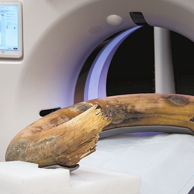
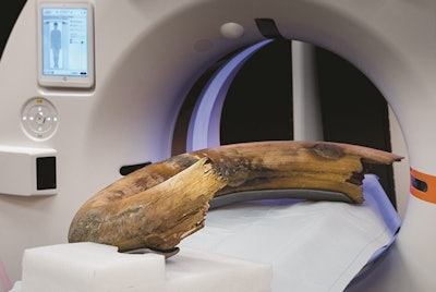
 Photograph of the positioning of the woolly mammoth tusk in the scanner. The tusk was fixed in a fiberglass frame to guarantee transport and table-movement stability. There was 1 cm of clearance within the gantry bore. Image courtesy of Radiology.
Woolly mammoths used their tusks for fighting, scraping bark off trees, and digging for food. The tusks consisted of a bone-like substance called cementum and tissue underneath the cementum called dentin; the dentin was stacked in cone-shaped cups that accrued throughout the animal's lifetime.
In collaboration with the Institute of Evolutionary Medicine at the University of Zurich, Niemann and colleagues used the larger-gantry CT scanner to image the fossil, finding 32 cones, which indicates that the mammoth was at least that age when it died approximately 17,000 years ago, according to the RSNA.
"It was fascinating to see the internal structure of the mammoth tusk," Niemann said in the RSNA statement.


