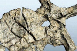
Dutch researchers used a CT scanner manufactured by Philips Healthcare to scan the fossilized tail vertebrae of a 66-million-year-old Tyrannosaurus rex dinosaur, using a spectral imaging protocol that provided researchers with new insight into the specimen.
A group at the Naturalis Museum in Leiden, the Netherlands used the company's IQon spectral CT scanner to examine the tail vertebrae of Trix, a T-rex specimen that was discovered in Montana, U.S. in 2013 and purchased by the museum. Trix was transported to Naturalis on 23 August.
Trix had been buried underground for 66 million years, and over time a mineralization process occurred that replaced her original bones with fossilized representations, with her tail vertebrae particularly well-preserved. A conventional CT scanner was previously used to scan the vertebrae, but a pyrite deposit in the tail blurred the images by absorbing x-rays.
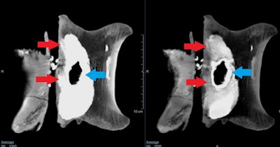
Instead, paleontologist Anne Schulp of Naturalis decided to use the IQon spectral CT scanner to scan four tail vertebrae (C20 - C23). By using the scanner's dual-layer detector and advanced spectral reconstruction algorithms, Schulp was able to able to obtain detailed images of the tail vertebrae, as indicated below.
 Comparison of a conventional CT image (left) with IQon image (right). The observed density in the conventional CT image is caused by the pyrite infill visible between the spinal canal and the main body of the vertebra, but is removed in the IQon spectral CT image. This reveals not only bone structure (red arrows), but also the structure of the pyrite infill (blue arrows).
The researchers noted these are just the first images acquired of Trix, and more are expected in the future.


