German researchers have reported at RSNA 2021 that volumetric assessment of bone mineral density on dual-energy CT (DECT) exams can yield accurate predictions for future risk of osteoporosis-associated fractures.
Researchers from Goethe University Frankfurt in Frankfurt am Main found that quantitative analysis of bone mineral density on DECT yielded an area under the curve (AUC) of 0.937. They believe that the technique could enable retrospective, opportunistic assessment of bone mineral density and enable high-risk patients to be treated before they have an osteoporosis-associated fracture.
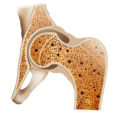
Osteoporosis is one of the most prevalent metabolic diseases; approximately 12.6% of the population over 50 years are affected by the disease in the U.S., according to presenter Leon Grünewald. And about one in three women and one in five men will suffer an osteoporosis-associated fracture in their lives.
At the same time, around 80 million CT exams are performed each year in the U.S., and a lot of the information available in the exams -- especially quantitative information -- isn't utilized, he said.
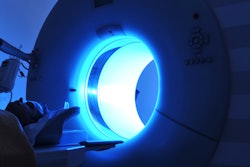
Quantitative CT methods do exist, but these techniques typically require calibration on phantoms, he said. However, dual-energy CT facilitates the acquisition of data on material decomposition, enabling differentiation of the materials of the bone and assessment of the density and the structures in the bone without requiring a phantom, Grünewald said.
"So this is really an ideal technology for the assessment of bone mineral density," he added.
In their study, the researchers sought to investigate whether retrospective and opportunistic bone-mineral assessment by dual-energy CT of the spine can actually predict the risk of osteoporosis or osteoporosis-associated fractures.
"The goal of osteoporosis diagnosis is that you can start treatment before any fracture happens," Grünewald said.
The researchers included 92 patients who had received dual-energy CT of the lumbar spine of the first lumbar vertebrae. Patients were excluded from the study if they had received an intravenous contrast agent; were suspected to have malignancy of the vertebral spine; were suspected or had actual inflammatory changes, degenerative changes, or fracture of L1; or had previous spinal surgery.
The study authors used dedicated DECT postprocessing software to manually delineate the trabecular bone on two image series. Bone mineral density was then extracted from a 3D region of interest.
They then performed receiver operating characteristic (ROC) analysis to obtain the optimal threshold for differentiating between patients who went on to have an osteoporosis-associated fracture in the next two years and those who didn't. The researchers found that the optimal threshold of 93.7 kilograms per meter yielded a high level of accuracy, as detailed in the chart below.
| Performance of dual-energy CT for identifying patients at risk for osteoporosis-associated fractures | |
| Sensitivity | 85.5% |
| Specificity | 89.2% |
| Area under the curve | 0.937 |
Grünewald noted that the threshold is actually higher than the American College of Radiology's recommendations for quantitative CT. The researchers believe that the higher threshold is due to the elimination of inaccuracies -- such as the fat area -- encountered with normal quantitative CT techniques. These errors can lead to an underestimation of bone mineral density, he said.
"And dual-energy [CT] actually eliminated this error," Grünewald said. "So I think we're probably closer to the real value of bone."
In the future, the researchers hope to develop an automated approach for bone mineral density assessment, as well as consider the use of contrast-enhanced exams.
"And we're also looking into locoregional measurements of other areas of the body that could actually also provide information for opportunistic assessment of bone mineral density," Grünewald said.
