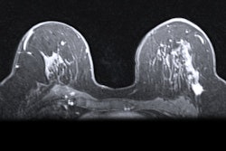
Contrast-enhanced mammography (CEM) may be an efficient diagnostic workup method for women undergoing breast cancer screening who have been recalled, suggest findings published on 3 July in the Lancet Regional Health Europe.
Researchers led by Dr. Marc Lobbes, PhD, from Maastricht University Medical Center in the Netherlands, found that CEM achieved a diagnostic accuracy comparable to that of conventional breast imaging. However, the results favor CEM’s use due to better efficiency in terms of resources used, as well as more occult lesions being detected.
“[Our study] showed that CEM is, in our opinion, the preferred modality to use after a screening recall,” Lobbes told AuntMinnieEurope.com.
Many women who are recalled from breast cancer screening receive post-screening work-up in the hospital with full-field digital mammography. However, other imaging methods such as digital breast tomosynthesis (DBT), MRI, and ultrasound can be used in a supplemental role to determine the final diagnosis, with or without tissue sampling. Radiologists have been trying to determine the best courses of action to mitigate false-positive cases, which may cause unnecessary anxiety and costs for women.
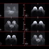
The Lobbes team presented findings from the RACER trial, a multicenter, randomized controlled clinical trial that compared CEM to conventional imaging (such as digital mammography or DBT) as a primary breast imaging modality in a real-world scenario. It also evaluated diagnostic workups in women recalled from breast cancer screening. Lobbes said that to the best knowledge of the researchers, this trial is the first of its kind.
Women in the trial were randomly assigned to either a CEM branch or a control branch for conventional imaging as a primary workup tool in two general and two academic hospitals.
The final analysis included data collected between 2018 and 2021 from 526 women. Of the total women, 264 were assigned to CEM while 262 were placed into the control branch.
| Comparison between CEM, conventional imaging in RACER trial | |||
|---|---|---|---|
| Measure | Conventional imaging | CEM | p-value |
| Sensitivity | 97.7% | 98% | 1 |
| Specificity | 75.4% | 75.6% | 1 |
Based on only primary full-field digital mammography/DBT or CEM, the final diagnosis was reached in 27.7% of women who underwent CEM and 1.1% of women in the control branch. Additionally, the conventional imaging branch demonstrated a significantly higher frequency of supplemental imaging compared with the CEM branch (p < 0.0001).
The team also noted that the median time needed to reach the final diagnosis was comparable between both cohorts at one day. This included a range of zero to four days for CEM and zero to three days for conventional imaging. Finally, CEM detected 13 malignant occult lesions versus three from conventional imaging, and no serious adverse events occurred.
Based on their results, the study authors suggested that CEM “should be strongly considered” as the preferred primary imaging modality in women recalled from breast cancer screening. They added that the risks tied to CEM use in this area are “negligible,” with Lobbes calling it a “safe and feasible” method.
“To complete the circle, we are currently performing a cost efficiency analysis as well, to study whether the approach with CEM is also cheaper,” Lobbes told AuntMinnieEurope.com.
The full study can be found here.