Low-dose iron oxide tracers impact the quality of breast MR images, but not that of contrast-enhanced mammography (CEM), a study published on 5 February in Surgical Oncology found.
Researchers led by Elisabeth van Haaren from the Zuyderland Medical Centre in Sittard-Geleen, the Netherlands, found that even after a low-dose injection (1 ml) of superparamagnetic iron oxide (SPIO) tracer, iron remnants stay behind in the breast tissue and disturb all MR images. However, they also observed no such effect on CEM images.
“CEM could be valuable alternative if additional imaging is needed in the follow-up of breast cancer,” van Haaren and colleagues wrote.
SPIO tracers are used to find and retrieve sentinel lymph nodes in clinically node-negative breast cancer patients. The researchers highlighted that this method is noninferior in performance compared to Tc99m-nanocolloid and blue dye. They added that this method is nonradioactive and thus avoids strict logistical guidelines in place for radioactive materials.
Previous studies suggest that iron oxide particles that remain in breast tissue after these tracers are used disturb MR images by presenting as susceptibility artifacts. Meanwhile, other studies have demonstrated that CEM further improves full-field digital mammography to the point where it may be considered noninferior to breast MRI.
The van Haaren team studied the effect of iron particles that remain after a low-dose injection of SPIO tracers on the quality of breast images from MRI and CEM. It focused on the first year of follow-up after breast-conserving treatment and sentinel lymph node biopsy.
The researchers included 14 women in their prospective study. The SPIO tracer was injected seven days before sentinel lymph node biopsy and wide local excision were performed. The women also underwent follow-up CEM and breast MRI consecutively, except for one who refused to undergo MRI. However, one other woman had a bilateral carcinoma, so SPIO was injected in both breasts.
Two radiologists meanwhile scored the quality of the images using a four-point Likert system: 0 (no artifacts), 1 (good diagnostic quality), 2 (impaired but still readable), and 3 (hampered clinical assessment).
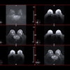
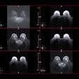
No CEM image in the study showed artifacts, meaning the radiologists issued a Likert score of 0 for all CEM images. However, the radiologists observed susceptibility artifacts caused by residual SPIO particles on all 14 breast MR images and issued a Likert score of 2.
The researchers also noted that the results were independent of the SPIO injection site, the artifacts occurred on all sequences, and they could still evaluate the contralateral breast.
They added that their results are concordant with those of previous studies evaluating both modalities in this area.
“The advantage of CEM lies in the fact that an ultrasound can be performed immediately as a ‘one-stop shop’ appointment and should be therefore preferred, since it reduces the number of hospital visits,” the study authors wrote.
The study can be found in its entirety here.


