Dear MRI Insider,
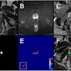
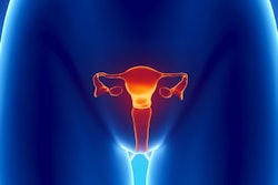
In a suspected case of cervical cancer, evaluating parametrial invasion can be essential to determine the most appropriate management of the patient and to improve outcome. By far the most accurate way of doing this is with T2-weighted imaging, according to prize-winning researchers from Italy.
The team from Modena won many plaudits for its groundbreaking study at ECR 2019. Admittedly the study only involved a small sample, but the key findings are well worth a read.
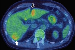
MRI has become increasingly popular for evaluating the various stages of prostate cancer and cases of its recurrence -- largely because of the modality's soft-tissue contrast and the emergence of diffusion-weighted imaging -- but PET/CT with the radiopharmaceutical gallium-68 (Ga-68) prostate-specific membrane antigen has won support too. German investigators have asked: Which technique is better?
Meanwhile, a French group has used a basic MRI method to measure iron content in the substantia nigra to obtain a better reading on how stroke affects the brain and long-term patient outcomes. Called R2* mapping, the technique can detect increased iron concentrations, and it can be added to follow-up MRI scans to monitor neurodegeneration and resulting poor motor function among stroke patients.
In Russia, the use and availability of MRI have come under scrutiny. A five-year "road map" has been produced for the Moscow region, and we've interviewed Prof. Sergey Morozov, PhD, about the implications.
Training in MRI is one of the areas being modernized by Dr. Nicola Strickland, president of the U.K. Royal College of Radiologists, and her team. Don't miss our video interview about her plans and priorities for the next six months.
This letter features only some of the many articles posted over the past few months in the MRI Community. Please scroll through the full list of our coverage below.

