A new overview article has offered not only insights into the use of ultrasound and MRI in assessing blood flow and vasculature of uterine fibroids but also a generic terminology for describing these parameters.
"Blood supply is crucial in the genesis, diagnosis, and treatment of fibroids," noted Derk Jan Slotman, PhD, of University Medical Center Utrecht, the Netherlands, and colleagues, in an article published in Insights Into Imaging on 16 September. "Ultrasound and MRI can unravel the complex vascular composition of fibroids."
 Sagittal fibroid images obtained by the different ultrasound techniques. 2D B-mode ultrasound (A); 2D power Doppler (B); 2D microvascular flow (C).All images courtesy of Derk Jan Slotman et al and Insights Into Imaging. Available for republishing under Creative Commons license (CC BY-NC-ND 4.0).
 3D power Doppler images of the same patient. Sagittal view (A), transversal view (B), and coronal view (C). 3D power Doppler reconstruction (D) with a 3D power Doppler volume (E) and a histogram (F) as output of 3D power Doppler ultrasound with vascular indices as result.
The authors' main aim was to describe the use of different imaging modalities suitable for assessing vasculature, blood flow, and tissue microstructure of uterine fibroids, giving detailed information on the blood supply of the fibroids, while using consistent terminology. They have provided a table as a glossary for their proposed nomenclature, listing each term with its definition. This consistent terminology is an important aspect of their review; until the present time, there has been no consistent generic nomenclature for the blood flow and vasculature of fibroids, they explained.
Consistent terminology is critical to avoid miscommunication between radiologists and referring doctors, they added. Their terminology is offered as a means of improving both interdisciplinary communication and clinical management of fibroids. This is particularly important as the blood flow and vasculature of fibroids are most often imaged with ultrasound or MRI. While the MRI interpretation is usually the task of radiologists, the authors note that ultrasound is often interpreted by referring doctors.
The authors observe that the two primary modalities are complementary, with ultrasound being employed as first-line imaging for assessment of the vasculature, blood flow, and tissue microstructure of fibroids. Furthermore, at the point of initial diagnosis, assessment of blood flow can discriminate fibroids from pathologies such as adenomyosis or sarcomas.
In the article, they discuss the range of ultrasound methods, including the different Doppler techniques used for perfusion imaging. Microvascular flow (MV) and contrast-enhanced ultrasound imaging can be used for blood flow in smaller blood vessels, according to the authors, who describe the benefits and limitations of each method.
Clinical use of MRI
MRI is used as the second-line modality when ultrasound cannot provide conclusive answers. "This may occur in selective cases when doubts exist regarding the benign/malignant nature of the supposed fibroid, or when MRI is necessary to assess eligibility for minimally invasive (uterine artery embolization) and non-invasive (MR-guided high-intensity focused ultrasound) treatments.”
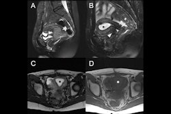
 MR angiography images of subserosal/intramural uterine fibroid with a diameter of 144 mm in the dorsal uterine wall and hypertrophic arteria overica in a 41-year-old woman (left), and subserosal pedunculated fibroid of a 42-year-old woman with a 69 mm diameter (right).
Tomographic 2D and 3D MRI may also provide critical information, with more anatomical details and better soft-tissue contrast, they added.
Overall, the article can assist readers in selecting the most useful modalities and techniques for future investigations of uterine fibroids as well as clinical practice, the authors suggest. The proposed common vocabulary should also be of value. “Clinical work and future fibroid studies focusing on angiogenesis, differentiation between benign/malignant, treatment selection or outcome prediction may benefit from uniform nomenclature as proposed in this work,” they concluded.
Read the full article here.