PET scans performed at one or three months after patients with large B-cell lymphoma begin chimeric antigen receptor T-cell (CAR T-cell) therapy can indicate whether they are responding to the treatment, according to a study published on 17 April in the Journal of Nuclear Medicine.
The finding could provide crucial insights for clinicians, enabling more personalized and timely interventions to improve patient outcomes, noted lead author Dr. Andrea Farolfi, of IRCCS University Hospital of Bologna in Italy, and colleagues.
“Noninvasive techniques such as F-18 FDG-PET/CT are needed to identify which patients are at the highest risk of [progressive disease], reduction in treatment duration, and death,” the group wrote.
CAR T-cell therapy has emerged as a groundbreaking treatment for patients with relapsed or refractory large B-cell lymphoma (LBCL), yet nearly half of patients experience disease progression or relapse within the first year after treatment, the authors explained.
Prior studies have found that parameters observed on F-18 FDG-PET/CT scans can predict how patients with lymphoma may respond prior to beginning CART T therapy, the authors noted. However, the best timing for scans for predicting responses after patients begin therapy remains in question.
Thus, in this study, the researchers investigated the prognostic value of PET parameters -- specifically maximum standard uptake value (SUVmax), total lesion glycolysis (TLG), and metabolic tumor volume (MTV) -- measured at one and three months after initiating CAR T-cell therapy.
The researchers analyzed images from 61 patients with LBLC who previously had undergone F-18 FDG-PET scans at different time points as part of a CAR T-cell treatment trial. During an 18-month follow-up period, 28 (46%) of the 61 patients had died.
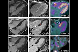
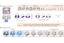
 A visual abstract of the study. Image available for republishing under Creative Commons license (CC BY 4.0 DEED, Attribution 4.0 International) and courtesy of the Journal of Nuclear Medicine.
According to a Kaplan-Meier analysis, SUVmax, MTV, and TLG were significantly associated with overall survival. Patients with an SUVmax of 6.3 or greater on PET scans at three months had an eightfold increase in risk of death (hazard ratio [HR], 8.15) compared with those below this cutoff. Similarly, higher MTV (≥120.1) on scans at three months yielded a nearly 10-fold risk of death (HR, 9.87).
In addition, SUVmax, MTV, and TLG on PET scans at both one and three months were associated with overall survival and progression-free survival (all p < 0.05) and PET scans at three months correlated with patients’ duration of response (p < 0.05).
“F-18 FDG-PET/CT at three months may offer slightly stronger prognostic discrimination, but both time points can be used for early risk stratification,” the group wrote.
Ultimately, the study reinforces a growing body of evidence suggesting that semiquantitative PET parameters offer valuable prognostic information beyond the traditional methods in assessing the response to CAR T-cell therapy, the authors wrote.
“Our findings support the incorporation of semiquantitative PET parameters derived one and three months after CAR T-cell infusion into routine clinical practice for assessing response to CAR T-cell therapy in patients with LBCL,” the group concluded.
The full study can be found here.