A new study has found that substantial differences in image quality exist in a range of hybrid imaging systems, even for similar clinical indications. This is due to variations in CT dose, linked to the CT scan parameters and applied CT reconstruction techniques, they say.
The data obtained can be used to establish image quality (IQ) reference levels in hybrid imaging as a tool for further optimization, noted Gwenny Verfaillie, a PhD researcher at Ghent University who also works as a medical physics expert in radiology at Jan Yperman Ziekenhuis, Ypres, and AZ West, Veurne, and her colleagues.
"Although the risks from PET/CT and SPECT/CT are generally far outweighed by the benefits, hybrid imaging results in increased radiation exposures due to the combined dose from the CT component and the radiopharmaceutical," they explained in an ECR 2024 poster presentation. "To optimize multimodality imaging, both image quality and dose need to be studied."
The CT acquisition may be performed for distinct reasons, and the image quality requirements differ according to the clinical task, which is reflected in the radiation dose to the patient. "For attenuation correction and anatomical localization of the emission data, the CT dose can be relatively small. For hybrid systems with diagnostic capabilities, higher exposure levels are more likely."
The dose from the radiopharmaceutical is related to the amount of administered activity, which depends on both the radiopharmaceutical used and weight of the patient. Due to improved hardware and software, image quality can be maintained at lower activities, the researchers pointed out.
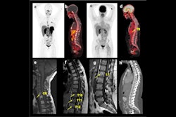
Objective inter-device comparisons of CT image quality are missing for the wide variety of PET/CT and SPECT/CT systems in use, and this prompted them to evaluate the CT image quality and corresponding CT dose of frequently performed examinations considering the clinical purpose of the CT scan. The group used the Catphan 440 phantom (The Phantom Laboratory), and to mimic the body of a standard adult patient, an elliptic polymethyl methacrylate (PMMA) annulus with a size of 20 cm by 30 cm was employed.
Using the open-source image processing software ImageJ, the group evaluated CT image quality by means of the contrast-to-noise ratio (CNR) and the spatial resolution (modulation transfer function, MTF). The CNR was investigated for the Teflon sensitometric target in the CTP401 Catphan module. It is defined as the ratio of the difference in mean signal, expressed in Hounsfield Units (HU), within a region of interest (ROI) placed in a target volume and in the background, and the background noise. To assess the spatial resolution, the MTF was calculated using the impulse created by the tungsten carbide located in the CTP592 module of the phantom.
This was done for the whole-body (WB) and bone protocols on three PET/CT (Siemens Biograph mCT Flow, GE Discovery MI, GE Discovery MI 3R) and four SPECT/CT (Siemens Symbia Intevo 6, GE Discovery NM/CT 670, Philips BrightView XCT, GE Infinia Hawkeye) systems. While the Philips BrightView XCT and GE Infinia Hawkeye are featured with a so-called dedicated low-dose CT, all other hybrid systems have a conventional CT with diagnostic capabilities. For each protocol, the CT scans were taken as in clinical practice. To study the relation with the CT dose, the displayed CT dose index volume (CTDIvol) was used.
Because image quality is influenced by the dose of the CT scan, the CTDIvol was used to compare the different clinical CT protocols and hybrid systems. As can be seen in the two figures below, large variations in CT dose were observed for both the WB PET/CT and bone SPECT/CT examinations.
 CT dose index-volume (CTDIvol) values of anatomical localization (AL-CT, shown in blue) and diagnostic (DI-CT) whole-body (WB) CT scans performed on three different PET/CT systems for the Catphan phantom. Note that for the Discovery MI 3R machine, no localization scans are performed clinically. All figures courtesy of Gwenny Verfaillie et al and presented at ECR 2024.
When looking at the hybrid imaging devices having a conventional CT, the diagnostic WB CT dose was about twice as high for the Siemens unit than for the GE PET/CT systems, while the dose for the localization CT was about three times smaller. For the SPECT/CT systems, the dose was higher for both the diagnostic and localization CT on the GE system. "These variations in dose are caused by differences in CT scan protocols for the same clinical indication, including the tube voltage, the use of automatic tube current modulation, the applied pitch, and collimation. Variations in CT reconstruction parameters, such as the employed reconstruction technique, also play a role," the authors noted.
 CT dose index-volume (CTDIvol) values of anatomical localization (AL-CT, shown in blue) and diagnostic (DI-CT) whole-body (WB) CT scans performed on four different SPECT/CT systems for the Catphan phantom. Note that the Philips Brightview XCT and GE Infinia Hawkeye have a dedicated low-dose CT without diagnostic capacities (T = thoracic, HQ = high quality, LD = low dose).
Applying a smooth reconstruction filter resulted in a CNR ranging from 24 to 55 and 80 to 98 for a localization and diagnostic WB CT, respectively. This variation in CNR is related to the used CTDIvol. A higher CT dose leads to less image noise, resulting in a higher CNR. For the diagnostic WB CTs reconstructed with a sharp kernel, both dose and specific kernel influence the CNR of 14-25. In bone SPECT/CT, conventional CT systems perform better than dedicated low-dose CT devices. Depending on the CT dose, CNR values range from 1 to 28 and 12 to 46 for, respectively, localization and diagnostic bone CT scans.
Overall, the spatial resolution is higher for diagnostic CT scans, and this is mainly due to the higher CT doses associated with these scans, the researchers said. "The applied reconstruction filter also affects the spatial resolution. Sharp filters namely aim to strengthen the edges and small details for imaging tasks that require more detail, such as bone scans."
On the other hand, smooth reconstruction filters help to reduce the image noise, and the spatial resolution also falls. This is the case for soft tissue imaging, where noise is more disturbing than blur. Similar results are observed for the three manufacturers -- Siemens, GE, and Philips -- and for both diagnostic and nondiagnostic CT exams. The spatial resolution is higher for images reconstructed with a sharp kernel.
The co-authors of this poster were Prof. Dr. Ir. Yves D'Asseler and Prof. Dr. Ir. Klaus Bacher. The project received funding from the Euratom research and training program.