FAPI-PET imaging could emerge as a new tool for assessing patients with single pulmonary tumors, especially in those with negative results on other scans, according to a study published on 11 April in the Journal of Nuclear Medicine.
A group at University Hospital Heidelberg in Germany assessed the approach in 19 patients referred by their treating physicians because of inconclusive lung cancer findings on CT and F-18 FDG-PET. FAPI-PET either confirmed or ruled out the disease in all cases, the group found.
“Our results suggest that supplemental Ga-68 FAPI-46 PET may improve the noninvasive assessment of primary pulmonary lesions compared with F-18 FDG-PET and CT alone,” wrote lead author Dr. Manuel Röhrich and colleagues.
CT and F-18 FDG-PET are considered clinical gold standards for lung cancer imaging, yet several subtypes of lung cancer, especially the lepidic subtype, are frequently negative on these scans. This “markedly hampers” the assessment of single pulmonary lesions, the authors wrote.
Conversely, PET imaging using gallium-68 (Ga-68)-labeled fibroblast activation protein (FAP) inhibitors has shown high potential for imaging various malignancies, and in this study, the group sought to assess its clinical value in these cases.
Between February 2022 and April 2023, 19 patients with suggestive pulmonary lesions underwent CT, F-18 FDG-PET, and Ga-68 FAPI-46 PET at the University Hospital Heidelberg. The cohort consisted of five female and 14 male patients between the ages of 41 and 77. All patients underwent CT and F-18 FDG-PET as clinical routine scans and were individually referred for additional imaging by their treating physicians because of inconclusive findings, the authors noted.
According to the results, Ga-68 FAPI-46 PET scans revealed whether primary lung tumors were malignant (n = 12) or benign (n = 7), with intense Ga-68 FAPI-46 radiotracer uptake seen in tumors in F-18 FDG-negative scans.
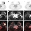
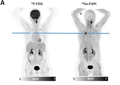 F-18 FDG and Ga-68 FAPI-46 images of a 51-year-old woman with adenocarcinoma with lepidic growth pattern in right upper lobe. (A) Maximum-intensity-projection PET images. The lesion had F-18 FDG uptake below blood pool level but was strongly Ga-68 FAPI-46-positive. CT-guided biopsy led to pathologic diagnosis of adenocarcinoma, and the patient was treated by stereotactic body radiation therapy because of functional inoperability. Image courtesy of the Journal of Nuclear Medicine.
“In our analysis, all cases of [lung cancer] showed markedly elevated Ga-68 FAPI-46 uptake, increased [tumor-to-background ratios], and increased Ga-68 FAPI-46/F-18 FDG ratios for all parameters compared with benign pulmonary lesions,” the group wrote.
In addition, the researchers performed a separate lab test on 24 tissue sections of lepidic lung cancer and found strong FAP positivity in all specimens. This characterizes lepidic lung cancer as a strong target for Ga-68 FAPI-46 PET imaging in F-18 FDG-negative cases, they suggested.
“This advance target characterization was of crucial interest for our analysis, as the presence of cancer-associated fibroblasts in lepidic [lung cancer] has already been described histologically but the FAP expression of this entity had not, to our knowledge, been evaluated before,” they wrote.
While the results are promising, additional research is warranted, the authors noted.
“Larger, confirmative studies are necessary to gain more evidence on the clinical value of Ga-68 FAPI-46 PET for assessment of primary pulmonary lesions,” the authors concluded.
Access to the full study is available here.


