Two new approaches to creating 3D images using x-rays could improve screening for diseases and the study of very fast processes, as well as enable analysis of material properties and provide structural information of opaque objects with unprecedented detail, an international research team has discovered.
One approach may reduce the x-ray doses needed for some types medical imaging, such as breast cancer screening. The other approach may allow 3D imaging of delicate biological samples or the study of very fast processes to speed development of more durable materials.
Led by Andrew Kingston of the Australian National University along with a team at the European Synchrotron Radiation Facility (ESRF) in France, the researchers demonstrated for the first time that the imaging approach known as ghost imaging can be used to obtain 3D x-ray images of the interior of objects opaque to visible light (Optica, November 2018, Vol. 5:12, pp. 1516-1520).
"Because of the potential for significantly lower doses of x-rays with 3D ghost imaging, this approach could revolutionize medical imaging by making x-ray screening for early signs of disease much cheaper, more readily available, and able to be undertaken much more often," said senior author David Paganin, from Monash University in Australia, in a statement. "This would greatly improve early detection of diseases, including cancers."
Ghost imaging correlates two x-ray beams that individually do not carry any meaningful information about the object. One beam encodes a random pattern that acts as a reference and never directly probes the sample, while the other beam passes through the sample.
The researchers created random x-ray patterns by shining a bright beam of x-ray light through metal foam. They took a 2D image of this random beam and then passed a very weak copy of it through the sample. A large-area, single-pixel detector captured x-rays that passed through the sample. The process was repeated for multiple illuminating patterns and sample-object orientations to construct a 3D tomographic image of the object's internal structure.
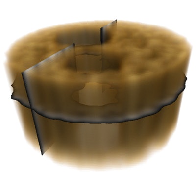
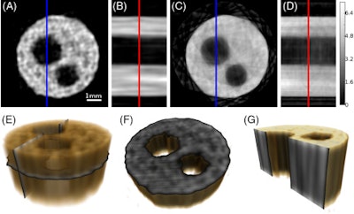 Horizontal (A) and vertical (B) 2D slices through the 3D x-ray GT reconstructed volume with a voxel pitch of 48 µm. The corresponding horizontal (C) and vertical (D) 2D slices through the conventional 3D tomography reconstructed volume obtained from the same set of experiments. (E) A semitransparent rendering of the 3D GT reconstructed object indicating the location of the slices (A) and (B). Horizontal (F) and vertical (G) cutaway images of the rendered ghost-tomogram volume showing the position of slices (A) and (B), respectively. Note that the blue lines in (A) and (C) indicate the position of the orthogonal slices (B) and (D); likewise, the red lines in (B) and (D) indicate the location of perpendicular slices (A) and (C). Image courtesy of Andrew Kingston et al, "Ghost tomgography, Optica, November 2018, Vol. 5:12, pp. 1516-1520.
As a proof-of-concept experiment, the group performed ghost x-ray tomography on an aluminum cylinder with a diameter of 5.6 mm and containing two holes smaller than 2.0 mm in diameter. The researchers were able to produce 3D images with 1.4 million voxels.
X-ray ghost imaging is a new field that needs to be further explored and developed, the study authors noted. "With more development, we envision ghost x-ray tomography as a route to cheaper and, therefore, much more readily available 3D x-ray imaging machines for medical imaging, industrial imaging, security screening, and surveillance," Kingston stated.
High-brilliance x-ray sources
In the other paper, a team from the Paul Scherrer Institute in Switzerland, led by Marco Stampanoni, and researchers from the Deutsches Elektronen-Synchrotron (DESY) in Germany and the ESRF, acquired 3D images using high-brilliance x-ray sources. Their new approach uses a single exposure to obtain 3D information from x-rays 100 billion times brighter than a hospital x-ray source. The rays can only be produced at specialized synchrotron facilities (Optica, November 2018, Vol. 5:12, pp. 1521-1524).
The technique can make the measurements necessary to create a 3D image before destruction of the sample, so it could be useful for studying the mechanics of delicate biological samples, such as examining the internal 3D structure of intact viruses or proteins.
The new single-shot approach uses a crystal to split one incoming x-ray beam into nine beams that simultaneously illuminate the sample. Using detectors oriented to record information from each beam allows researchers to acquire at once nine different 2D projections of a sample object before it is destroyed by the intense x-ray probe beams.
The researchers used the approach to image a moth, which demonstrated the potential for studying insect mechanics with 3D microscale resolution at speeds ranging from microseconds to femtoseconds. They also showed they could achieve nanoscale resolution by imaging a gold nanostructure.
The researchers plan to use their single-shot multiprojection imaging technique to better understand insect biomechanics, which could inspire new engineering setups. They also want to study new, lighter materials that might lower fuel consumption for vehicles, and plan to examine the fast processes that occur when space debris hits satellites, which could aid development of protective materials.

