To ensure timely diagnosis and treatment of ovarian torsion in emergency settings, clinicians must recognize its signs on CT imaging, according to research presented at the recent Royal Australian and New Zealand College of Radiologists (RANZCR) meeting.
Dr. Azri Johari of the radiology department of Mackay Hospital and Health Services in Queensland described case series findings regarding this uncommon but emergent condition that involves twisting of the ovarian vascular pedicle.
Ovarian torsion represents approximately 2.7% to 3% of all acute abdominal pain presentations in women, and its nonspecific presentation can make diagnosis challenging, as the symptoms can mimic other acute abdominal conditions such as appendicitis or renal colic, the authors wrote. Delayed diagnosis and treatment of the condition may result in venous and lymphatic obstruction with subsequent potential for arterial compromise and infarction, they noted.
Furthermore, while Doppler ultrasound is the first-line exam for diagnosis of ovarian torsion, it has technical limitations, especially with ambiguous findings, the team added.
Johari and colleagues explained that CT, which is often used when patients present with abdominal pain in emergency departments, has a demonstrated sensitivity for ovarian torsion ranging from 74% to 95% and specificity from 80% to 90% when characteristic features are present, in contrast to ultrasound’s respective sensitivity and specificity of 80% and 88%.
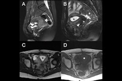
 CT coronal; 25-year-old woman -- a large cystic mass in the anterior mid pelvis with smooth hyperdense borders. It converges to the right at a focal area of vascular twisting (whirlpool sign).All figures courtesy of the Medical Imaging Department, Mackay Base Hospital, Mackay, QLD, Australia, and the RANZCR 2025 annual scientific meeting.
The case series not only “reinforces the importance of cross-sectional imaging in the emergency setting, but also the importance of recognizing key CT features of ovarian torsion to ensure timely and effective patient management,” the investigators wrote.
In both cases presented in the poster, the diagnosis of ovarian torsion was made using CT; this diagnosis was confirmed with timely surgical intervention for both patients. While the patients’ symptoms varied, both involved nonspecific clinical findings with persistent pain, which led to contrast-enhanced CT exams of the abdomen and pelvis.
One patient, a 25-year-old woman, presented with right-sided tenderness and pain with nausea and vomiting over two days. She was afebrile, hemodynamically stable, and her lab markers were within normal ranges. The other patient, a 37-year-old woman, was also hemodynamically stable and afebrile; her tenderness and pain were left-sided and had persisted for four days.
 CT sagittal; 25-year-old woman -- the cystic mass is causing mass effect on the urinary bladder and uterus. The left ovary is displaced posteriorly behind the mass (not imaged on this slice).
Large ovarian cystic masses were present in both cases; the authors underscored that these are a known risk factor for ovarian torsion, and thus an important finding for diagnosis.
In the first case presented, CT showed a large right-sided adnexal cystic mass with evidence of a twisted vascular pedicle referred to as the “whirlpool sign,” suggestive of ovarian torsion. CT in the second case also showed a large lesion, which appeared to originate from a left-sided pedicle.
 CT coronal; 37-year-old woman -- a large cystic mass in the anterior mid pelvis with smooth hyperdense borders. It converges to the right at a focal area of vascular twisting (whirlpool sign).
CT imaging showed key features in both cases that facilitated surgical intervention and treatment, the authors wrote, which underscores the important role the modality has in the time-sensitive diagnosis of ovarian torsion.
Read the full poster here, on the EPOS section for the RANZCR ASM 2025, available on the European Society of Radiology's website.