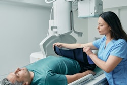
The application of an out-of-plane shield in CT scanning may lead to a substantial increase in tube current modulation, therefore providing patients with a much higher radiation dose, researchers from Finland have found.
"Shielding may have unintended effects on patient dose in CT," noted Heli Larjava, a specialist medical physicist, and her colleagues at Turku University Hospital. "Applying an out-of-plane shield outside the scanned volume but visible in the localizer images may increase the patient dose considerably if the scanner's tube current modulation function is based only on localizer images."
There are two types of contact shielding: bismuth shields placed on the patient and inside the imaged volume (in-plane shields), and lead shields wrapped around the patient, outside the imaged volume (out-of-plane shields). The authors' study, which was published on 14 September in European Radiology, focused on out-of-plane shields.
Using an anthropomorphic phantom, they evaluated the effect on tube current modulation of the out-of-plane shield visible in the localizer but absent in the scan range in chest CT with six different CT scanners from three different vendors.
The team first scanned the chest without any shielding, and then with the out-of-plane shield within the localizer but outside the imaged volume, using all pitch values of each scanner. The tube current values with and without the out-of-plane shield were collected and used to evaluate the effect of overscanning and tube current modulation on patient radiation dose.
The highest increase in cumulative mA was 217%, when the pitch was 1.531. The tube current value increased already 8.9 cm before the end of the scanned anatomy and the difference between the tube current of the last slices (with and without the out-of-plane shield in the localizer) was 976%.
Features like overscanning may be difficult for the user to notice when planning the scanning, and yet they may affect tube current modulation and through it to patient dose, the team pointed out.
Implications of study
Overall, the results were in line with expectations, Larjava told AuntMinnieEurope.com. "However, with some scanners, the huge difference in the end of scans (with and without the out-of-field shield) was a surprise. We were not expecting to see over ninefold difference in mA of the last slices of the scan."
She hopes that the results will have an impact on the shielding debate, particularly helping technicians who are unsure whether to use a shield in CT scanning.
"It is not easy in the clinical situation to decide such a thing, especially if the patient is demanding that the shield is used," she continued. "The local medical physicist should check all of the scanners, if the hospital wishes to use out-of-field shields -- there may be notable differences between them. In our hospital, there was already a policy of not using the out-of-field shield, but from time to time it was questioned."
Larjava would be very keen to see the performance of Philips scanners, which the researchers were unable to include in their research. "Also, the performance of the Canon scanners had changed completely from the older to the newer model -- it would be interesting to see whether there is a similar change to be seen with other manufacturers."
Clinical reality
The study confirms previous publications and advice against including lead shielding within the CT localizer, given the reliance on automatic exposure control on the localizer attenuation information, Shane Foley, PhD, associate professor and head of radiography at University College Dublin, told AuntMinnieEurope.com.
"While many professional groups now advise against any use of shielding due to various factors such as increased risk of overexposure from AEC impact, artifact creation and image quality degradation etc, recent work from Granata et al shows shielding is still in relatively wide use in Europe -- therefore reinforcing the need for those who continue to use it to be aware of the associated risks and in CT to ensure the shield is only placed after the localizer -- if at all," he noted.
Also, it is well known that the CT pitch has an impact on the overscan range, reminding users to use pitch wisely, especially in some radiosensitive cohorts, Foley stated, adding that it would have been very interesting to know if image artifacts were noticed and at what point/position were artifacts introduced due to the impact of the shield on the helical interpolation (as part of overscanning).
"What does surprise is the absolute radiation dose increases noted here," he said. "Admittedly the authors removed the maximum mA level, but a 900% dose increase is significant, despite the fact the lead was distant from the scan range and that indeed this area could well include radiosensitive breast and lung tissue," he commented.
You can read the European Radiology article here. The supervisor, Dr. Hannele Niiniviita, is also assistant chief medical physicist (diagnostic radiology) of medical physics department. The other co-author was Chibuzor Eneh. The study was supported by the Radiology Society of Finland and the State Research Funding of the Turku University Hospital.


