Zero echo-time (ZTE) is shining light on a wide range of developmental, traumatic, inflammatory, rheumatological, and oncological conditions and can open the door for more MRI-based musculoskeletal (MSK) studies and morphometric analyses, researchers from a leading U.K. hospital have reported.
"One MRI examination with a ZTE sequence allows cross referencing of sequences, aiding diagnosis, prognostication, and surgical guidance in soft tissue and bone with precise measurements that involve bony landmarks," said Helen Prince, senior MRI radiographer and MSK lead at Addenbrookes, part of the Cambridge University Hospitals National Health Service Trust, U.K.
"It brings promising possibilities in routine patient services and research," she reported in an International Society for Magnetic Resonance Technicians (ISMRT) poster session held on 2 June at the International Society for Magnetic Resonance in Medicine (ISMRM) annual meeting in Toronto.
Key benefits of ZTE
ZTE is an MRI sequence that visualizes tissues such as bone with the shortest T2 values giving images with a CT-like appearance, Prince explained. Signal is acquired immediately after applying the radiofrequency pulse, resulting in a near zero echo time, with the next radiofrequency pulse following in a very short repetition time.
"This rapid switch from transmit to receive mode enables acquisition of quickly decaying signal starting at a near zero echo time, capturing what little signal is present, especially in cortical bone," she continued. "Absence of on and off gradient switching gives a virtually silent acquisition."
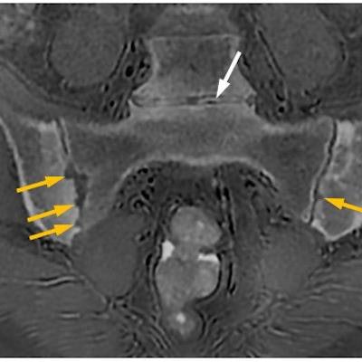
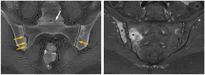
 A 39-year-old woman with ankylosing spondylitis. Erosions surrounding the sacroiliac joints (yellow arrows), which are structural lesions pointing to chronic damage, are conspicuously revealed on the CT-like ZTE MR image (left), whereas the STIR MR image (right) superbly shows periarticular osteitis (asterisks) consistent with active sacroiliitis. White arrow showing the L5-S1 intervertebral disk space on the left image points to a pitfall of the ZTE sequence: Gas within joint spaces and soft tissues gives the false appearance of calcifications. Nevertheless, MRI with the ZTE sequence that produces CT-like images affords one-stop imaging in many musculoskeletal conditions obviating the need for additional ionizing radiation-based examinations. Figure courtesy of Üstün Aydingöz, MD, Hacettepe University, Ankara, Turkey.
In theory, zero echo time could help to visualize MR imaging of any joint, according to Prince. ZTE sequences are resilient to artifacts caused by motion and magnetic field homogeneities, with great signal-to-noise ratio and scan time efficiencies, and they have advantages in depicting small bone fragments in a trauma setting and subtle cortical erosion.
"ZTE may remove the need for CT, with detailed depiction of bone anatomy, making an MRI examination a one-stop imaging examination for conditions such as femoroacetabular impingement, giving both soft tissue and bone imaging within the same examination," she noted. "Substantial agreement is being found in morphometric analyses, with reproducibility of many lytic or sclerotic lesions -- although spatial resolution is still inferior to CT or radiography."
It has shown value in bone morphology, assessment of fractures, displaced bone fragments, shoulder instability, calcification (ossification of ligaments, calcific tendinitis) spinal foramina stenosis, skull assessment (suture closure, trauma), and assessment of bone erosions in bone tumors, Prince added. It may also demonstrate a wide range of structural abnormalities and disease or healing processes.
What are the pitfalls?
An important pitfall is that the spatial resolution of MRI ZTE 3D reformats is not adequate to aid surgical planning, so patients are still undergoing CT scans, she cautioned. "The reduced acoustic noise in comparison with the rest of the exam means that some patients move, thinking that the scan is complete."
The drawbacks of ZTE are readily offset by cross-checking with other MRI sequences. Correlation with other standard MR images is essential for correct characterization of misleading signal intensity on ZTE such as hemosiderin deposition, gas, and microscopic fragments within joints or tissue mimicking calcification or ossification, Prince stated.
In spite of these difficulties, ZTE has been implemented clinically at Addenbrookes, where radiology staff conduct an average of 36,000 MRI exams a year on six scanners from GE Healthcare.
For further information, she recommends the following article: Aydingöz Ü. Zero Echo Time Musculoskeletal MRI: Technique, Optimization, Applications and Pitfalls. 2022, Radiographics 42, 1398-1414.


