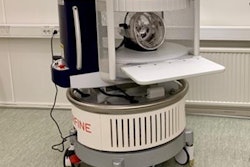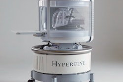
A prototype low-cost, low-field MRI scanner operating at 0.055 tesla could expand access to neuroimaging in low- and middle-income countries, according to a study published on 14 December in Nature Communications.
Its use could also translate into a better patient imaging experience by mitigating noise during scanning and removing the need for radiofrequency shielding, wrote a team led by Yilong Liu, PhD, of the University of Hong Kong in Pokfulam.
"Together with complete elimination of radio frequency and magnetic shielding cages, our ultra-low-field MRI approach can offer a more patient-friendly alternative [to high-field MRI]," the group noted.
MRI is a valuable diagnostic tool for evaluating brain injury and disorders, providing noninvasive, nonionizing, quantitative, and multiparametric imaging. Yet despite these advantages, only 30% of the world's population has access to MRI, the authors wrote.
There are seven MRI scanners per 1 million people around the world -- compared to 200,000 CT scanners and 1.5 million ultrasound scanners -- and most of these are concentrated in high-income countries. Then there's cost and upkeep: MRI scanners can cost between $1 million (8.8 million euros) to $3 million (2.6 million euros), with monthly maintenance of $15,000, and they have high power requirements to keep the magnets cool.
The investigators developed a compact, mobile, low-cost, low-field 0.055-tesla MRI device that they estimate could be produced for about $20,000 (17,650 euros) and operates from a standard AC wall outlet with no radiofrequency or magnetic shielding. They also developed a "deep-learning driven EMI [electromagnetic interference] cancellation technique to model, predict, and ... remove the external and internal EMI signals from MRI signals, thus eliminating the traditional radio frequency shielding cage," they wrote.
The team used the low-field scanner to assess 25 patients for neurological disease, including stroke and tumors. Scan protocols included T1- and T2-weighted imaging, fluid-attenuated inversion recovery (FLAIR), and diffusion-weighted imaging. Total scan time was 30 minutes. Patients also underwent a 3-tesla MRI exam using standard protocols (scan time, 20 minutes).
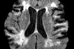
The prototype MRI scanner identified most key pathologies in all 25 patients, compared with the conventional 3-tesla scanner: In one brain tumor case, both the 0.055-tesla and conventional MRI images showed a mass in the right parietal cortex that was "hypointense in T1W and hyperintense in T2W," the group wrote. The study also demonstrated the following:
- T1W images on both types of MRI scanners showed clear asymmetry between the right and left hemisphere, indicating a cystic lesion.
- The prototype scanner showed "excellent sensitivity and correspondence" with the conventional scanner in identifying a choroid plexus cyst at the occipital horn of the right lateral ventricle.
- Both types of scanners identified hematoma on a subacute intracranial hemorrhage case.
- The 0.055-tesla scanner showed "little susceptibility" to artifacts from metallic clips and stents compared to the high-field device.
This prototype scanner offers benefits such as open magnet configuration, low noise levels during scanning, low sensitivity to metal implants, and fewer image artifacts, and shows promise for expanding access to MRI, the authors concluded.
"We succeeded in implementing four essential standard clinical imaging protocols on this [device] ... and demonstrated the preliminary clinical feasibility in diagnosing tumor and stroke," they wrote. "The development of such an ultralow-field MRI technology will enable patient-centric and site-agnostic MRI scanners to fulfill unmet clinical needs across various global healthcare sites and has the potential to democratize MRI for low- and middle-income countries."


