An experimental radiotracer based on gallium-68 (Ga-68) can help identify primary and metastatic tumors on hybrid PET/MRI scans in women with advanced breast cancer -- even those smaller than 1 cm, according to a study published on 12 October in Radiology.
A German group from the University of Muenster analyzed the performance of a Ga-68-labeled fibroblast activation protein inhibitor (FAPI) PET tracer (Ga-68 FAPI-46) on whole-body PET/MRI scans for detecting primary and metastatic tumors in women with advanced breast cancer. They found the tracer is a promising new option for helping diagnose the disease.
"[Ga-68 FAPI-46] PET/MRI showed strong and reliable accumulation in invasive cancer ... and provided contrast even for the detection of subcentimeter lesions," wrote first authors Drs. Philipp Backhaus and Matthias Burg.
F-18 FDG is the most frequently used tracer for breast assessment. F-18 FDG is used for whole-body staging in advanced breast cancer but has limited accuracy in evaluating primary breast lesions due to low uptake levels, according to the authors.
Fibroblast-activation protein (FAP) is abundantly expressed in various cancers, and studies suggest it is a more specific target for imaging tracers. Ga-68 FAPI-46 was first synthesized and evaluated in early studies for detecting cancer by a German group at the University of Heidelberg in 2019 and is currently being studied in several clinical trials.
"But results in primary breast tumors remain lacking," the authors wrote.
In this study, the researchers retrospectively analyzed 18 patients with large or locally advanced breast tumors who underwent PET/MRI scans (Biograph mMR, Siemens Healthineers) after Ga-68 FAPI-46 injections between October 2019 and December 2020 at University Hospital Muenster.
All patients had histologically confirmed breast cancer, and most had larger or locally advanced breast tumors. Standardized uptake values (SUVs) indicating the amount of tracer accumulated in tumors were identified with dedicated software.
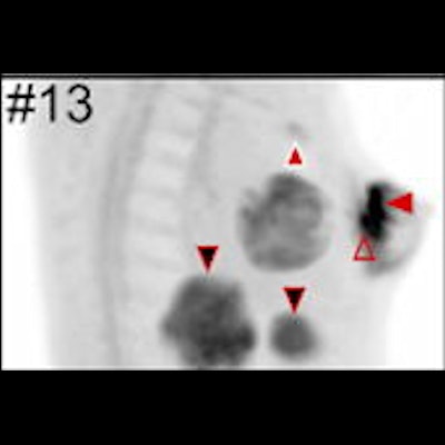
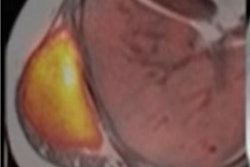
The researchers observed strong tracer accumulation in all 18 untreated primary breast malignancies. Clear tumor delineation was seen across different gradings, receptors, and histologic types, with a mean maximum SUV of 13.9 (range, 7.9 to 29.9) and a median lesion diameter of 26 mm (range, 9 to 155 mm). Separate studies have determined mean SUVmax uptake of FDG in primary breast cancer tumors is 5.7.
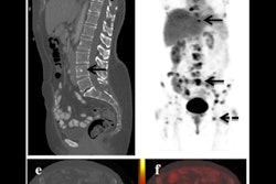
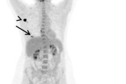
In addition, all preoperatively verified lymph node metastases in 13 women showed strong tracer accumulation (mean SUVmax = 12.2). Tracer uptake established or supported extra-axillary lymph node involvement in seven women and affected therapy decisions in three women, the researchers reported.
"This retrospective analysis indicates promise for gallium-68 fibroblast-activation protein inhibitor tracers for breast cancer diagnosis and staging," the authors concluded.
 Fibroblast-activation protein inhibitor breast PET/MRI scans. Oblique coronal projections of prone breast PET/MRI maximum intensity projection images in all 18 patients who underwent breast PET/MRI. Image intensities are adjusted to standardized uptake value on a scale of 0-10. Image courtesy of Radiology.
In an accompanying editorial, Drs. David Mankoff, PhD, and Mark Sellmyer, PhD, of the University of Pennsylvania, rued the researchers' choice of PET/MRI versus the more widely used PET/CT. MRI may hold advantages for imaging primary tumors, yet studies comparing PET/MRI and PET/CT for breast cancer nodal and distant disease staging are limited. In these settings, no studies to date show major advantages of PET/MRI over PET/CT, they stated.
Overall, however, the research shows considerable promise for the application of Ga-68 FAPI-46 PET in breast cancer for staging and possibly for primary breast cancer diagnosis, the authors wrote.
"This early report supports future studies to explore both applications with a focus on tumor types and clinical scenarios where FAPI PET may provide key clinical data and insights that are not provided by current clinical imaging approaches," Mankoff and Sellmyer concluded.


