Skier's thumb represents between 5% and 10% of all skiing injuries, but skiing actually only accounts for 3% of acute ulnar collateral ligament (UCL) injuries. It can pose a serious diagnostic challenge, but one that can be overcome if six top tips are kept in mind, according to a leading musculoskeletal (MSK) imaging expert.
"Ultrasound is an excellent technique to image skier's thumb, provides high resolution images, and has shown to be highly accurate with surgical correlation," noted Dr. Catherine McCarthy, consultant MSK radiologist at Oxford MSK Radiology and Nuffield Orthopaedic Centre, Oxford, U.K., during a session on full-thickness tears of the UCL at the recent virtual ultrasound workshop hosted by the European Society of Musculoskeletal Radiology (ESSR).
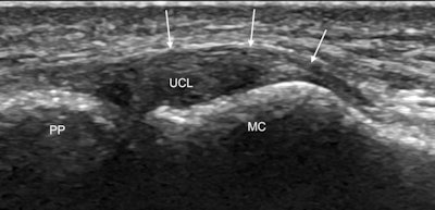 Coronal ultrasound image along the ulnar aspect of the thumb MCP joint shows the normal UCL as a convex echogenic fibrillar band (UCL) bridging the joint between the metacarpal head (MC) and first proximal phalanx (PP). The adductor aponeurosis is seen as a thin curvilinear echogenic structure (arrows) overlying the UCL. All images courtesy of Dr. Catherine McCarthy.
Skier's thumb typically occurs during a fall, when the ski pole forces the thumb into radial abduction, and it is an injury of the UCL of the metacarpophalangeal (MCP) joint. Dynamic imaging with flexion and extension of the interphalangeal joint should be used for identifying key anatomy in skier's thumb injuries, while comparison with the asymptomatic side will help confirm diagnosis, McCarthy said.
The following are her six main recommendations for imaging tears:
- Know the normal anatomy and the spectrum of UCL injuries.
- Distinguish undisplaced tears from Stener lesions, as this determines management: An undisplaced tear is symmetrically centered over the MCP joint, whereas a Stener lesion has an asymmetrical nodular appearance centered on the metacarpal head.
- Optimize ultrasound parameters, transducer type, and patient position.
- Learn the ultrasound signs of normal ligament and aponeurosis, as well as the three patterns of full thickness tears.
- Deploy dynamic imaging with flexion and extension of the interphalangeal joint to identify anatomy.
- Compare sides to confirm diagnosis.
Lesions such as avulsion or an undisplaced intrasubstance tear of the ulnar collateral ligament usually heal spontaneously. However, in about a third of cases, the torn ligament retracts proximally and displaces superficial to the adductor pollicis aponeurosis. This injury is known as a displaced tear or Stener lesion, and the patient must undergo surgical repair to prevent permanent instability and premature osteoarthritis, McCarthy explained.
It isn't always easy clinically to distinguish between an undisplaced tear and a Stener lesion, so ultrasound diagnosis is vital for determining patient management, she noted. For such a diagnosis, optimizing ultrasound parameters is key.
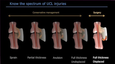 Radiologists must be familiar with the spectrum of ulnar collateral ligament injuries.
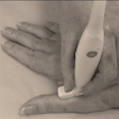
First, the patient should sit opposite the sonographer with the palm of the hand and radial aspect of the thumb flat on the examination table, allowing access to the ulnar collateral ligament. Given the superficial location of the ulnar collateral ligament, sonographers should use a high frequency 18 MHz small footprint hockey stick transducer as it allows easy positioning into this small anatomical space.
"You should initially place the transducer in the longitudinal plane and look for a small concavity on the metacarpal head," McCarthy noted.
Ultrasound signs
Spotting the concavity seen on the metacarpal head, between the metacarpal tubercle and the articular surface, is key to identifying the normal ulnar collateral ligament and confirms that the transducer is correctly oriented at the ligament attachment. The ulnar collateral ligament should appear as an echogenic fibrillar band bridging the MCP joint. The deeper fibers are predisposed to anisotropy, and this should not be confused with a partial thickness tear, McCarthy stated.
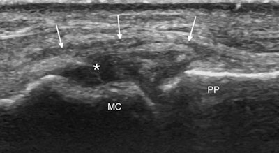 Coronal ultrasound image along the ulnar aspect of the thumb MCP joint of an undisplaced UCL tear. The ligament is diffusely thickened and hypoechoic with a cleft extending through its proximal aspect (asterisk). The ligament remains symmetrically centred over the MCP joint and deep to adductor aponeurosis (arrows).
The next structure to look for is the adductor aponeurosis, which can be seen as a thin curvilinear echogenic structure overlying the ulnar collateral ligament. It may be difficult to identify the adductor aponeurosis in the static position so she advocates the use of dynamic imaging with flexion and extension of the interphalangeal joint to move the aponeurosis which will then be seen as a thin echogenic band gliding over the ulnar collateral ligament.
Tear identification
In an undisplaced tear in which the ligament looks thickened and hypoechoic often with a cleft extending through it, there is no significant proximal retraction, so the ligament appears symmetrically centered over the MCP joint. Dynamic imaging once again using joint extension and flexion will show the aponeurosis gliding over the thickened hypoechoic torn ligament. Meanwhile, in Stener lesions the torn ligament retracts proximal to the MCP joint to lie adjacent to the metacarpal head. The ligament has an asymmetrical appearance with a larger retracted proximal end, which looks like a round hypoechoic mass.
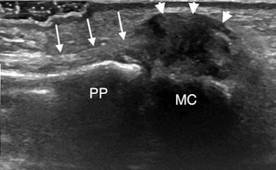 Coronal ultrasound image along the ulnar aspect of the thumb MCP joint demonstrates a displaced UCL tear or Stener lesion. The proximally retracted UCL appears as a heterogeneous mass (arrowheads) adjacent to the metacarpal head (MC) which is displaced proximal and superficial to the adductor aponeurosis (arrows).
"The adductor aponeurosis will point into the retracted ligament and does not overlie it. This nodular appearance recalls the 'yo-yo on a string' sign visible at MRI with the aponeurosis making up the string extending into the retracted nodular ligament, which forms the ball of the yo-yo," McCarthy said. "You may also see the aponeurosis folding between the fibers of the torn ligament which are displaced superficial to the aponeurosis."
Again dynamic imaging will show the aponeurosis clashing into the retracted ligament that is displaced proximal and superficial to the aponeurosis, and this identification method should be preferred to radial stress. It is often useful to compare with the normal asymptomatic side to help clarify the picture and confirm the diagnosis, while keeping an eye out for an avulsion fracture, she concluded.


