A new diffusion MRI scoring scale could improve diagnosis of the prion malady sporadic Creutzfeldt-Jakob disease (sCJD) and compares favorably to cerebrospinal fluid testing, according to a study published on 1 June in JAMA Neurology.
A team led by Dr. Alberto Bizzi of Fondazione IRCCS Carlo Besta Neurological Institute in Milan found that the new criterion increased diffusion MRI's sensitivity for identifying autopsy-confirmed sCJD compared with current criteria. The scale also made the modality similar in performance to cerebrospinal fluid testing, which is also used to diagnose prion diseases.
"[Our] findings suggest that diffusion MRI is an accurate test for establishing a diagnosis of prion diseases in the appropriate clinical context," the group wrote.
Diffusion MRI has long been used to diagnose prion diseases, with a basic protocol that includes identifying abnormal signals in the caudate and putamen, the researchers noted. In recent years, evaluation criteria for diffusion MRI for prion disease diagnosis were expanded to include signal intensities in the cortex of at least two brain lobes (with the exception of the frontal lobes on diffusion-weighted MRI and fluid-attenuated inversion recovery, or FLAIR); these updated criteria have now become the current standard.
But they may still be too limited, according to Bizzi and colleagues.
"These criteria might be too conservative, missing the diagnosis of patients with frontal cortex and/or only one lobe involvement or with only the caudate or putamen affected," the team cautioned.
Bizzi's group sought to evaluate the sensitivity and specificity of diffusion MRI for diagnosis of Creutzfeldt-Jakob disease with an index text that includes the new criterion of at least one positive brain region among the cortex of the frontal, parietal, temporal, and occipital lobes; the caudate; the putamen; and the thalamus. The investigators compared this new index test performance with current criteria, as well as with the performance of cerebrospinal fluid tests.
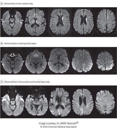
 A: A man in his 70s with sCJD subtype VV2 with signal hyperintensity in the body of the left caudate, without evidence of signal abnormality in the putamina and neocortical ribbon. B: A woman in her 60s with sCJD subtype MM1 with signal hyperintensity in the cortical ribbon of both parietal lobes, left greater than right, and left precuneus without evidence of abnormality in the striatum, thalami, and cortical ribbon of the temporal and occipital lobes. C: A woman in her 70s with sCJD subtype MM1 with signal hyperintensity in the precuneus (right greater than left) and right parietal cortex. Images courtesy of JAMA Neurology.
The study included diffusion MRI scans from 872 patients. Of these, 619 had sCJD, 102 had other prion diseases, and 151 had nonprion disease. Four radiologists scored the MRI scans of 200 patients randomly selected from the pool of 872; of the 200, 150 had sCJD and 50 had nonprion diseases.
The researchers found that when compared with current criteria, diffusion MRI using the newly proposed criterion improved specificity (90% to 95% versus 69% to 76%). It also made MRI's performance comparable to cerebrospinal fluid testing -- a particularly promising result, since MRI is noninvasive and more accessible.
| Cerebrospinal testing vs. diffusion MRI for diagnosing sCJD | ||
| Measure | Cerebrospinal testing | Diffusion MRI (using new criteria) |
| Accuracy | 87.5% | 95.5% |
| Sensitivity | 86.1% | 94.9% |
| Specificity | 100% | 100% |
The study findings could translate to improved patient outcomes, according to the authors.
"With the development of new potential therapies to alter the natural history of prion disease, early diagnosis is essential to maximize the chance of salvaging brain tissue," they concluded. "It is important that practitioners are aware of the diagnostic potential of diffusion MRI because it is the most widely available diagnostic test."


