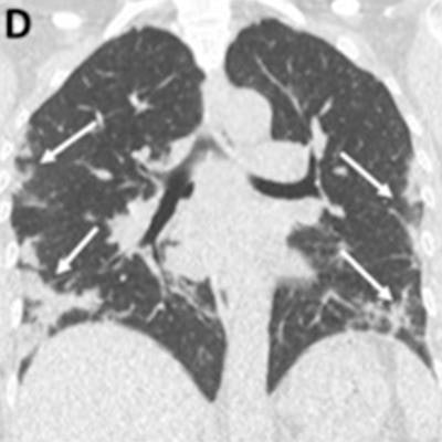
The Moscow Diagnostics and Telemedicine Center has made available for download a dataset of more than 1,000 sets of chest CT scans of patients with image findings of COVID-19.
To date, this is the largest cache of CT scans from patients with the SARS-CoV-2 virus. It can be used to develop artificial intelligence-based services.
All studies contain special markings that reflect manifestation of pathological abnormalities of COVID-19 in lung tissue based on the scans. Marking those areas of interest may lead to the development of artificial intelligence algorithms that can help radiologists locate suspicious areas on CT scans. Marking the pathology contouring can be used for automatic quantitative assessment of lung lesions and for comparing two CT studies of a patient.
Also, 50 of the studies were marked explicitly in patients where zones of ground-glass opacities and consolidations in pixels that are specific for COVID-19 are indicated on each CT slice with lung tissue abnormalities.
“The additional advantage of this dataset is that all CT scans included there were performed in primary healthcare facilities for the adult population. Besides that, it has been posted in public domain, and thin CT slices of up to 1 mm have already been converted into NIFTI format recognized among machine learning professionals,” said Dr. Sergey Morozov, chief regional radiology and instrumental diagnostics officer of Moscow Department of Health and CEO of the Diagnostics and Telemedicine Center in Moscow.
The database, which also contains 3D CT studies, includes studies that were collected between 1 March and 25 April using the Unified Radiological Information Service (URIS), which is connected to diagnostic equipment at 80 healthcare facilities in Moscow.


.png?auto=format%2Ccompress&fit=crop&h=167&q=70&w=250)


