Significant advances are occurring in placental MRI -- particularly the development of texture analysis, machine learning, and radiomic approaches -- and the time is now ripe for systematic and detailed MRI scans of the placenta in pregnancy, a leading French expert believes.
"Understanding how the placenta works is one of the major challenges facing radiologists, given the crucial role of this organ in fetal development," noted Prof. Nathalie Siauve, director of Diagnostic Radiologie Explorations Fonctionnelles Anatomopathologie Médecine Nucléaire (DREAM) at the Assistance Publique-Hôpitaux de Paris (AP-HP) Nord at the University of Paris.
Because radiologists now play a central role in detecting birth defects, adjusting clinical management, and establishing the prognosis of affected pregnancies, they must be aware of the new methods for evaluating the placenta so they can alert clinicians rapidly in cases of suspected abnormalities and facilitate appropriate timely management of the mother and the fetus, she wrote in a guest editorial posted online on 7 August by European Radiology.
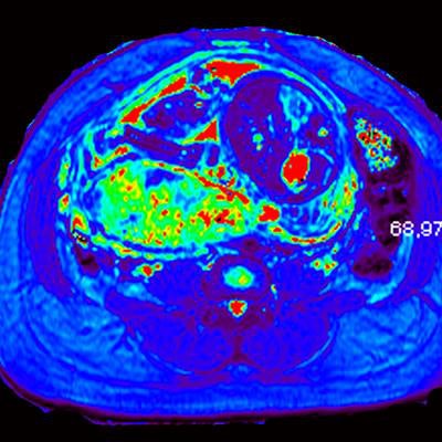
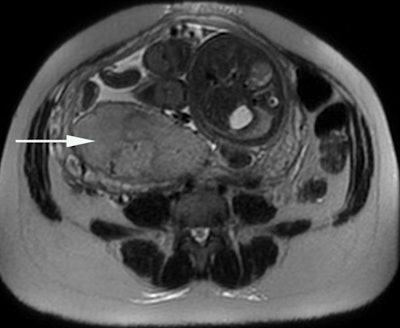 Placental oxygenation can be assessed by the blood oxygen level-dependent (BOLD) MRI technique, using T2* mapping (below) and combining a morphological analysis (above). The placenta is localized posteriorly on the right (arrow). The T2* value within each pixel is encoded with a color scale. Images courtesy of Prof. Nathalie Siauve.
"This organ plays a regulatory role extending well beyond nutrition and respiration, also encompassing the endocrine and immune system regulations," Siauve pointed out. "As a signaling organ, the placenta produces a myriad of bioactive molecules affecting both maternal and fetal metabolisms and physiologies."
The development and maturation of the placenta during pregnancy is relatively unknown, and it's time to harness modern approaches and technologies and to develop new ones to shed light on placental structure, development, and function in real-time, she emphasized. This is a priority of the U.S. National Institute of Child Health and Human Development's Human Placenta Project.
Historically, placental MRI was seen as a complementary problem-solving tool for placental evaluation, and it was much less common than fetal MRI, Siauve explained. However, placental abnormalities are of considerable clinical significance, due to their association with high rates of fetal morbidity and mortality, and placenta previa, placental adherence abnormalities, and placental insufficiency are key defects here.
"MRI has recognized added value for the management of placental adherence abnormalities, which requires a multidisciplinary team approach," she continued. "The early detection of placental adherence abnormalities is of crucial importance for determining the most appropriate surgical management technique and preventing hemorrhage during the delivery."
Novel perspectives
New avenues are opening up for MRI studies of the placenta. For instance, the early screening of women with a high risk of developing placental insufficiency by texture analysis may become feasible in the near future. Researchers have described textural changes in vivo in the placenta on MRI, in healthy and high-risk fetuses, and in the setting of placental insufficiency, they have demonstrated large differences in placental development relating to the onset and severity of fetal growth retardation and neonatal outcome, according to Siauve.
Texture analysis provides quantitative evaluation of heterogeneity through the quantification of gray-level patterns and pixel interrelationships within an image, and the textural features for each image are extracted from several matrices, she added. Therefore, machine learning and artificial intelligence identify the most relevant textural features and can be used to construct an optimal model to facilitate diagnosis.
Such an approach is particularly suitable for analyses of the placenta, which is known to be heterogeneous, Siauve wrote. This heterogeneity increases significantly with fetal aging during gestation, through both cotyledon maturation and other aging-associated processes, such as fibrin accumulation and calcification.
"Intensity heterogeneity is an emerging MRI marker of placental invasion. However, the reading of the images is subjective and this heterogeneity is difficult for human readers to quantify," she stated. "Texture analysis could be used for quantitative image analysis to assess placental heterogeneity. Machine-learning analysis with MRI-derived texture analysis features is a potentially feasible tool for the identification of placental tissue abnormalities underlying placental adherence abnormalities."


