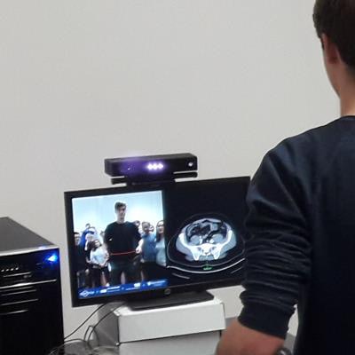
Augmented reality (AR) technology capable of displaying MRI and CT scans allowed researchers from Germany to offer a new, interactive learning method to medical students -- improving their understanding of anatomy and radiology, according to an article recently published in Anatomical Sciences Education.
The technology is based on a previous proposal by Blum et al to use the concept of a smart mirror -- a mirror-like screen that displays digital information -- to project AR models onto a person's body. This "AR magic mirror" consisted of a motion-sensing device (Kinect One, Microsoft) mounted on top of a split-screen monitor that displayed an MRI or CT scan on one screen and the same scan superimposed on a real-time video of the user on the other.
Through hand gestures, users were able to change the type of image displayed, switch between different image section planes (from axial to sagittal, for example), and modify aspects such as the contrast of CT scans. In addition, the AR magic mirror allowed users to examine an entire medical image volume within seconds simply by waving their hand up and down.
In the new study, researchers led by Daniela Kugelmann, PhD, of Ludwig-Maximilians University in Munich explored the potential of the AR magic mirror to facilitate anatomy and radiology education.
The AR magic mirror potentially offers a new look at anatomy and radiology education -- one that supports the shift toward interactive, multimodal learning environments and may help increase students' engagement and motivation, the authors noted.
To put the idea to the test, Kugelmann and colleagues offered the AR magic mirror as well as the Anatomage virtual dissection table as supplemental educational tools to 749 first-year medical students taking an anatomy course at their institution from 2016 to 2017 (Anat Sci Educ, 29 January 2019).
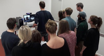 Medical students using augmented reality magic mirror during an anatomy and radiology course. Image courtesy of Felix Bork, graduate student at the Technical University of Munich.
Positive feedback on the usefulness of the AR magic mirror and Anatomage encouraged the researchers to offer both tools, in addition to traditional anatomy atlases, to medical students who signed up for a follow-up anatomy and radiology course. The students were divided into three groups, and each group used just one of the different learning tools for the entirety of the follow-up course.
On average, the students with access to the AR magic mirror received a higher score on the imaging portion of the post-test than did those who used the Anatomage and traditional textbooks and atlases. Using the AR magic mirror also resulted in the greatest improvement in scores at 35.3%, compared with 30.3% with the Anatomage and 28.7% with standard atlases.
| Comparison of different methods for anatomy and radiology education | ||||||
| Pretest | Post-test | |||||
| Standard atlas | Anatomage virtual table | AR magic mirror | Standard atlas | Anatomage virtual table | AR magic mirror | |
| Mean score for image-based questions | 30.4% | 28.8% | 29.6% | 59.1% | 59.1% | 64.9% |
After completing the follow-up course, the students filled out a survey in which they universally agreed that the AR magic mirror exceeded the Anatomage in terms of usability and benefits. Their responses also indicated that both methods could be effective supplemental teaching devices for radiology learning but could not necessarily replace traditional texts.
"AR systems for integrated radiology teaching in gross anatomy such as the magic mirror offer the potential to become a unique and powerful learning tool as well as an integral part of both modern medical curricula and students' educational toolsets," the authors wrote.


