Congenital agenesis of the pericardium is often discovered as an incidental finding, but Greek researchers have now demonstrated that CT and MRI can reveal important information about this rare cardiac malformation.
 Dr. Daphne Theodorou, PhD, is head of the MRI-CT unit at General Hospital "G. Hatzikosta" in Ioannina.
Most patients are asymptomatic, particularly those with complete absence of the pericardium, and physical examination and chest x-rays tend to be unremarkable. Echocardiography can provide useful information, but in many cases, CT or MRI is needed for subsequent confirmation.
"Chest radiographs may reveal levoposition of the heart, elevation of the cardiac apex, and elongation and flattening of the left border with prominence of the main pulmonary artery, which is separated from the aortic knob by interposed lung tissue," explained Dr. Stavroula Theodorou and Dr. Daphne Theodorou, PhD, radiologists at General Hospital "G. Hatzikosta" in Ioannina.
"CT and MRI can efficiently show the abnormal displacement and rotation of the heart and readily depict absent pericardium, as well as associated cardiac and extracardiac anomalies," they added in an e-poster presentation at this month's U.K. Radiological and Radiation Oncology Congress in Liverpool.
Clinical presentations
Congenital agenesis of the pericardium varies from complete absence of the left, right, or both sides of the pericardium to small regional defects. Partial defects may involve areas of the left, right, or diaphragmatic pericardium, according to the authors.
Absence of the pericardium allows interposition of lung tissue between the aorta and pulmonary artery, and, occasionally, bulging of the left atrial appendage through the defect or between the diaphragm and the base of the heart.
This unusual anomaly occurs in around 1 in 14,000 necropsies, and symptoms may include precordial pain, palpitations, dyspnea, syncope, and sudden death.
"Timely diagnosis can be missed owing to the rarity of the disease as clinicians or radiologists may not come across even a single case in their practices," the authors noted. "Nearly 30% of the patients with congenital agenesis of the pericardium have additional anomalies, including atrial septal defect, bicuspid aortic valve, mitral stenosis, tricuspid insufficiency, patent ductus arteriosus, tetralogy of Fallot, pulmonary sequestration, bronchogenic cyst, and congenital diaphragmatic hernia."
Significant learning case
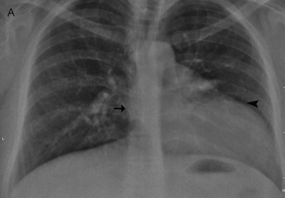
In one case the authors encountered at their hospital, an otherwise healthy 35-year-old woman was referred for ovarian cyst surgery. A chest radiograph demonstrated marked displacement of the cardiac silhouette to the left with flattening and elongation of the left ventricular contour (Snoopy sign) and obliteration of the right cardiac border (see figure).
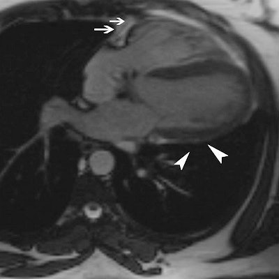
 Above A: Frontal chest radiograph shows displacement of the cardiac silhouette to the left, with flattening and elongation of the left ventricular contour (arrowhead) and obliteration of the right cardiac border (arrow). Below B: Gradient-recalled echo axial MRI shows complete absence of the left pericardium (arrowheads), with obliteration of pericardial fat and a partial right-sided pericardium (arrows) seen as a dark, thin stripe sandwiched between the epicardial and pericardial fat. Images courtesy of Dr. Stavroula Theodorou and Dr. Daphne Theodorou, PhD.
A cardiac MRI scan was performed to investigate cardiac levoposition and other associated abnormalities. Axial gradient-recalled echo (GRE) MR images showed a partial right-sided pericardium, seen as a dark stripe between the epicardial and pericardial fat, and complete absence of the left pericardium with obliteration of pericardial fat.
"On the corresponding coronal GRE MR images, there was interposition of lung parenchyma between the descending aorta, the diaphragm and diaphragmatic surface of the heart," they noted. "Diagnosis of congenital agenesis of the pericardium was instituted. No treatment was required as surgery is reserved for symptomatic patients and those at risk of sudden death due to apical ventricular herniation/strangulation through the pericardial defect."
Take-home messages
Agenesis of the left pericardium is the most common type, presenting in around 70% of cases, the authors wrote. Congenital agenesis of the pericardium occurs from premature atrophy of the common cardinal vein (duct of Cuvier), compromising blood supply to the left pleuropericardial fold, and it usually involves the left side.
"It is very important to differentiate partial from complete absence of the pericardium because only patients with partial agenesis of the pericardium are at risk of fatal myocardial herniation and entrapment of a cardiac chamber," they emphasized. "Surgical closure or enlargement of the defect (pericardioplasty) is occasionally required to prevent herniation."
Although in most cases, congenital agenesis of the pericardium is asymptomatic, accurate diagnosis is critical due to the risk of complications and sudden death in patients with pericardial defects, they concluded.
The other authors of the e-poster were Dr. S. Kakitsubata and Dr. Y. Kakitsubata, from the department of radiology at Miyazaki Konan Hospital in Miyazaki, Japan.


