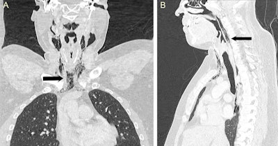
A CT scan confirmed that a man experienced a spontaneous perforation of his pharynx after attempting to stop a forceful sneeze, according to a new case report published online in BMJ Case Reports on 15 January.
A previously fit and well 34-year-old man presented to the emergency department with an acute onset of odynophagia and change of voice after a forceful sneeze, according to the authors from the University Hospitals of Leicester in Leicester, U.K. After he tried to halt a sneeze by pinching his nose and holding his mouth closed, the man experienced a popping sensation in his neck and some bilateral neck swelling.
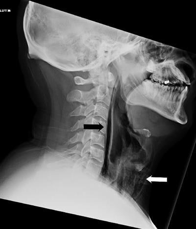 Lateral soft-tissue radiograph showed streaks of air in the retropharyngeal region (black arrow) and extensive surgical emphysema in the neck anterior to the trachea (white arrow). All images courtesy of BMJ Case Reports.
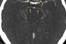
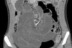
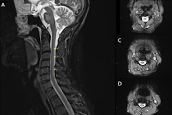
A lateral soft-tissue neck radiograph was performed, followed by an urgent, contrast-enhanced CT of the neck and thorax that confirmed the presence of extensive soft-tissue emphysema and pneumomediastinum.
After being admitted and treated conservatively with prophylactic intravenous antibiotics and enteral feeding via a nasogastric tube, the patient's symptoms and subcutaneous emphysema gradually resolved during the course of admission, according to the authors.
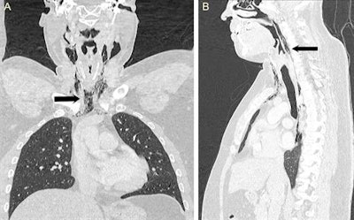 Coronal and sagittal CT scan of the neck and thorax confirmed the presence of surgical emphysema within the neck as well as pneumomediastinum extending from the skull base up to the T9 vertebra (black arrow).
"CT scan of the neck and thorax with water-soluble contrast swallow should be used as the gold-standard investigation, which can confirm the diagnosis and defines the exact pathological site," they wrote. "In addition, the normal CT appearance of the lung parenchyma and esophagus helps to exclude more serious causes of pneumomediastinum such as tracheobronchial rupture and Boerhaave's syndrome."
The full case report can be found here.


