In May of this year, a research team at the University Medical Center Utrecht (UMCU) in the Netherlands performed the first patient treatment using a new MRI-guided radiotherapy system that integrates a diagnostic quality 1.5-tesla MR scanner with an advanced linear accelerator. Now the team has published details of the first four patient treatments, demonstrating the feasibility and clinical utility of the MRI-linac system.
The Utrecht team treated four patients with lumbar spinal bone metastases with a single fraction of 8 Gy prescribed to the target volume while minimizing dose to the spinal cord and the rest of the body. The treatments demonstrated the system's ability to deliver precisely targeted radiation doses while simultaneously capturing the high-quality MR images that will allow clinicians to visualize tumors at any time, and adapt the treatment accordingly.
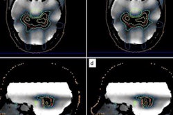
The patients were treated with a 3- or 5-beam step-and-shoot intensity-modulated radiotherapy (IMRT) plan. Plans were created while the patient was on the treatment table and based on the online 1.5-tesla MR images, with pretreatment CT deformably registered to the online MRI to obtain Hounsfield values (Physics in Medicine and Biology, 14 November 2017, Vol. 62:23, pp. L41-L50).
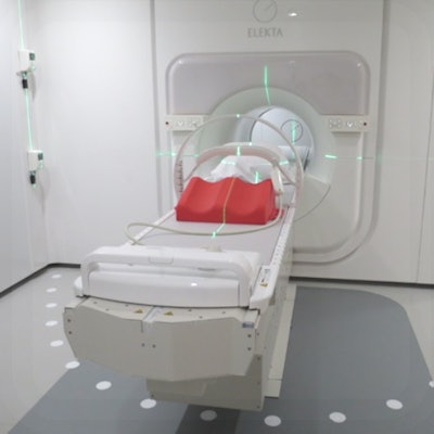
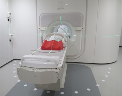
 The MRI-linac at the University Medical Center Utrecht, showing the lasers for patient set-up, the patient positioning devices and the radiofrequency receive coil.
"These study results are very promising and we look forward to further advancing the clinical development of this transformative system, which has the potential to revolutionize the treatment of cancer," said Bas Raaymakers, PhD, professor of experimental physics at UMCU. "These first clinical treatments show an outstanding level of dosimetric and geometric accuracy of the online IMRT planning and the radiation delivery based on the 1.5-tesla MRI guidance. This approach enables the optimization of dose to the tumor while reducing exposure of healthy tissue. To date, achieving this optimization has been the key challenge in radiation therapy."
The team chose bone metastases as the first treatment site as these tumors can be clearly visualized on MRI while the surrounding spine bone can be detected on the integrated portal imager. In this way, portal images can be used to independently verify the MRI-based guidance and quantify the precision of radiation delivery.
They validated the geometric accuracy of online MRI guidance by comparing portal images of the IMRT segments with the MRI-based calculated projections. This revealed an average beam alignment of 0.3 mm with the target as defined on the online MRI, demonstrating the stereotactic geometric system accuracy. For each patient, the team performed the same quality assurance procedures for both the pretreatment plan and the delivered plan, finding a mean difference of 0.4% of the calculated dose.
"These preliminary results are exciting and support the tremendous potential of Elekta's MR-linac (Unity) to address some of the historic challenges to improving the safety and efficacy of radiation therapy," commented Kevin Brown, global vice president of scientific research at Elekta. "The exceptional dosimetric and geometric accuracy reported in this study support the system as a transformative approach to radiation therapy that may allow more patients to receive optimum cancer care."
© IOP Publishing Limited. Republished with permission from medicalphysicsweb, a community website covering fundamental research and emerging technologies in medical imaging and radiation therapy.


