Using a combination of microbubbles, advanced image acquisition techniques, and signal processing, researchers at Imperial College London have a plan to extend ultrasound further into cancer diagnosis and treatment. The 42-month project, supported by Cancer Research UK (CRUK) and the Engineering and Physical Sciences Research Council (EPSRC) in the U.K., brings together long-term collaborators Mengxing Tang, Eric Aboagye, Nick Longand, and Dr. Adrian Lim -- an engineer, a biologist, a chemist, and a clinical radiologist.
"Building on our previous ultrasound work, the idea is to take what is a relatively affordable technology -- compared with PET and MRI choices -- and raise its capability in terms of sensitivity, specificity, and image contrast," Aboagye told medicalphysicsweb.
Taking advantage of ultrasound's system costs together with its point-of-care accessibility, the team is considering a wide range of applications, from detection and diagnosis through to cancer characterization, treatment design, and monitoring. "If we can target tumor-specific properties, then we can take ultrasound to a whole different level," Tang added.
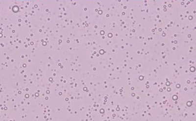
 Microbubble ultrasound contrast agents under the microscope.
To succeed, the scientists need to advance the technology on a number of fronts. And the multiyear funding is an essential element in their approach, allowing the group to examine each component in detail, including the contrast agent.
"One of the challenges is to make a bubble that is going to stick to a factor expressed selectively in the tumor," Aboagye said. "And we're able to take a step back and first address issues such as the batch-to-batch variation, characterizing every step of the bubble manufacturing process."
The bubbles serve as a highly sensitive contrast agent and have multiple uses. Furthermore, their similar size to blood cells allows them to stay within the tumor vasculature. "Without any targeting, they can show tissue function in terms of both macro- and microblood flow," Tang explained.
Image processing
Nine months into their work program, the researchers are also busy developing new hardware and software to enhance on-screen images. "Ultrafast ultrasound acquisition generates a huge amount of data, which invites advanced signal processing techniques," Tang said. "We are also looking at the design of the pulse sequences to further optimize the image acquisition."
Their goal is to translate this large pool of information into ultrasensitive detection, examining how different parameters can be tuned to extract different details across a range of imaging scenarios.
"Already, we have developed some imaging sequences and signal processing tools, which have allowed us to generate promising initial results in vivo," Tang said.
These early data are allowing the group to fast track key elements of the engineering and push ahead in refining the microbubble chemistry. In year two, the scientists begin the biology program, bringing the work closer to its final application.
"At the end of the project we will have the clinical development of ultrafast ultrasound, together with the preclinical development and understanding of the bubbles in vivo," Aboagye said.
The group faces numerous hurdles along the way, but is confident in its approach, highlighting the need to establish a close collaboration, especially for new teams. "I can imagine there might be more challenges if you had team members coming together for the first time," Aboagye said. "Success comes when you have the ability to regroup and make decisions quickly."
And it's not just cancer treatment that stands to benefit from the group's achievements. The planned upgrades in ultrasound imaging, particularly the improved temporal response of the setup, could find applications in cardiology -- an area the team is also targeting with its research.
The full title of the project is "3D ultrafast ultrasound with microbubble contrast agents for simultaneous imaging of molecular targets and perfusion in cancer."
Editor's note: This article is the fourth in a series looking at some of the research funded through the CRUK-EPSRC Multidisciplinary Project Award.
© IOP Publishing Limited. Republished with permission from medicalphysicsweb, a community website covering fundamental research and emerging technologies in medical imaging and radiation therapy.


