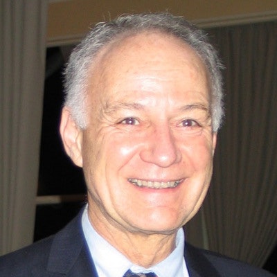
Dr. David Cosgrove, a leading researcher and teacher from London, died on Tuesday, 16 May, at the age of 78. An enthusiast for clinical ultrasound, microbubble contrast agents, and elastography, he remained active right up to his death.
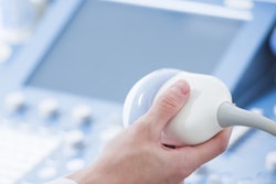
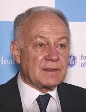 Take ultrasound more seriously and set aside more time for training, Dr. David Cosgrove recommended. Image courtesy of ET Healthworld.
Cosgrove, who was emeritus professor of clinical ultrasound at both Imperial College of Science, Technology and Medicine, and King's College, shared his views about the modality in a broad-ranging video interview recorded in January 2016 at the 69th Annual Conference of Indian Radiological and Imaging Association (IRIA), where he was the organizers' main VIP guest.
"Ultrasound's a strange subject in a way -- a lot of radiologists, for example in the U.S.A., don't regard ultrasound as an important discipline at all, yet it's the most widely used imaging technology in the world," he noted. "It's exploding and usage is being taken up very, very strongly, particularly in the less-well-off parts of the world, where it's become the standard."
Cosgrove said he was fascinated by what's happening in China, where ultrasound is not carried out by radiologists but by so-called ultrasonologists, who are medical doctors specializing in ultrasound and undertaking all types of examinations, including intervention, cardiac, and obstetrics. The government realized early on that ultrasound is a very cost-effective modality and invested heavily in it, he explained.
Unshakable belief in training
During his long career, Cosgrove was a strong advocate for training.
"Every imaging discipline needs good training, but in ultrasound there's a special aspect because it's such a practical technique," he said in the same IRIA video interview. "In a way, it's quite like a physical examination of the patient, say of the abdomen or the nervous system. The person doing the examination has to physically touch the patient with the probe and create the images as they're working with the patient in front of them."
There's great physical skill involved; it can't be taught quickly and requires many years of practice and hands-on training, added Cosgrove, whose main areas of expertise were the clinical role of nonobstetric ultrasound, the basic mechanisms of the ultrasound image-forming process and of Doppler, the applications of microbubble echo-enhancing (contrast) agents for ultrasound, and the application of elastography to clinical diagnosis.
"Ultrasound's quite a treacherous discipline. When it's done by an expert, it looks really easy, but it's not that easy to learn," he explained. "Take ultrasound more seriously and set aside more time for training so we can use it well for our patients."
Ultrasound is very safe at the powers currently in use, but it must be remembered that when administering ultrasound energy to the patient, physical changes are taking place. Keep the power levels down, particularly in high-intensity focused ultrasound, and pay attention to the regulations, he advised.
Career highlights
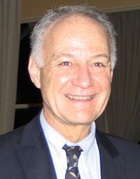 Cosgrove was involved in the early clinical development of elastography.
During the 1960s, Cosgrove undertook his training at St. George's Hospital Medical School in London and Oxford University, before completing his MSc degree in nuclear medicine from University of London in 1975, focusing on a comparison of liver scintiscans and ultrasonography in clinical diagnosis.
Between 1977 and 1993, he was a consultant in nuclear medicine and ultrasound at the Royal Marsden Hospital in London. He was professor of clinical ultrasound at Imperial College from 1994 to 2005, before becoming an emeritus professor. Cosgrove has also worked as a consultant radiologist at Hammersmith Hospital, London, and was a member of the scientific board of SuperSonic Imagine.
Cosgrove was involved in the preparation and publication of a series of guidelines into the clinical uses of contrast that helped lead to the U.K. National Institute for Health and Care Excellence’s guidelines on the role of contrast-enhanced ultrasound in characterizing focal liver lesions. He also headed up the preparation and publication of European guidelines on the clinical uses of elastography.
In his curriculum vitae (CV) posted on the focaltherapy.org website in 2011, he listed the following as his career highlights:
- First reports of clinically significant ultrasound findings, e.g. the features of biliary tree dilatation, pneumobilia, hemangiomas, abdominal tumors of various types, thyroid diseases, fatty changes in the liver, the use of Doppler in breast diagnosis, and transit time analysis of microbubbles in tumors.
- Reports on the mechanisms of ultrasound appearances (e.g. artifacts, such as transdiaphragmatic echoes) and on exploring novel means to extract hitherto unavailable information from the ultrasound signals. Generally known as tissue characterization, this work was carried out in close collaboration with the department of physics at the Institute of Cancer Research, particularly with Dr. Jeff Bamber and colleagues. These studies provided insight into the extreme complexity of the interactions between ultrasound and tissue and may yield practical applications (e.g. speckle reduction, induced motion analysis, or elastography). Doppler studies have focused on the clinical evaluation and introduction of techniques such as color and power Doppler.
- Echo enhancing agents became the major focus of study, with the establishment of a research team to investigate this unique opportunity both from fundamental and clinical points of view. Fundamental studies include nonlinear imaging and quantification of the change in echogenicity with microbubble concentration leading to functional indices and imaging. Clinical studies included phase III trials with a range of microbubble agents (especially in the liver and in tumors) and functional studies (especially in diffuse and focal liver diseases and in tumors).
Tributes from colleagues
"He was a fantastic and patient teacher and trained a whole generation of sonographers and radiologists. He was generous with his time and always made you feel valued," said Dr. Adrian Lim, adjunct professor of imaging at Charing Cross Hospital in London, and consultant radiologist at Imperial College. "He was a great mentor and over the years became a close friend, providing invaluable advice and support. We will miss him tremendously but cherish all the moments we were able to spend with him."
Similar sentiments were expressed by Dr. Chris Harvey, a consultant radiologist at Hammersmith Hospital.
"He was a very genuine, caring, and thoughtful person who was well liked and respected by his colleagues, and is a great loss to radiology," he said.
Funeral arrangements
The funeral ceremony will take place on Friday 2 June from 12:15 to 13:45 in the chapel at the South London Crematorium, which is within the Streatham Park Cemetery, Rowan Road, Streatham in London.
