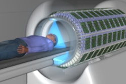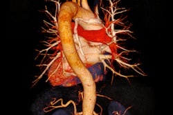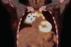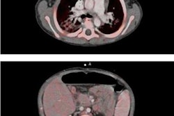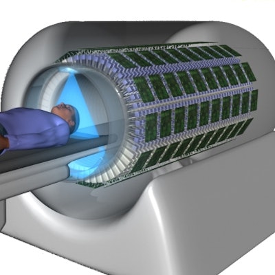
The EXPLORER PET system is a 2 m long, total-body scanner that could vastly increase the range of PET applications. Under development by the multi-institutional EXPLORER consortium, the scanner provides 30 to 40 times better sensitivity than current scanners. This allows high-throughput scanning, with the potential to perform a whole-body exam in a single breath-hold, and injected radiation doses as low as 10 MBq, enabling PET imaging of pediatric patients.
The consortium is currently constructing the first EXPLORER prototype. The final device will contain over 560,000 scintillator crystals and form over 90 billion lines of response (LOR) -- about 100 times more than current clinical PET scanners -- making data handling a major challenge. To address this task, the researchers developed a quantitative image reconstruction method for EXPLORER and used Monte Carlo simulations to evaluate the quality of the resulting images.
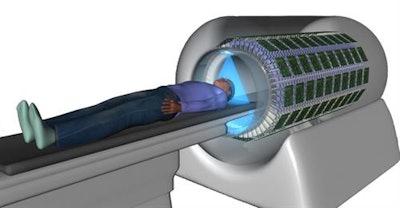 Artistic illustration of the EXPLORER system.
"Delivery of the EXPLORER prototype is expected in 2018," explained Simon Cherry, PhD, professor of biomedical engineering at the University of California, Davis, U.S., and co-leader of the EXPLORER consortium.
Image reconstruction
Because EXPLORER has a vast number of LORs, and records time-of-flight (TOF) information, the team chose list-mode data processing for 3D image reconstruction, as it is more efficient than sinogram-based reconstruction. The long axial field-of-view (FOV) can result in axial resolution loss, due to the parallax effect between crystal pairs in rings with a large axial spacing. To reduce this blurring effect, they applied a model estimated from point source data to compensate resolution loss in the reconstruction (Physics in Medicine and Biology, 27 February 2017).
The long FOV also makes EXPLORER more susceptible to multiple scattering than conventional PET scanners. The team used a Monte Carlo-based scatter estimation to model and correct for the occurrence of multiple scatters. They also estimated and corrected for randoms, where two detected photons arise from different annihilation events.
Scanner simulations
The researchers performed Monte Carlo simulations of a 20 minute PET scan on an anthropomorphic adult male phantom after injection of 25 MBq F-18 FDG -- about 20 times lower dose than the clinical standard. They generated time-activity curves for 14 major organs and tissues, plus lesions in the liver and lung.
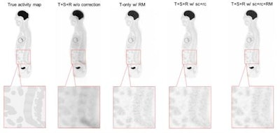 Reconstructed sagittal images with different levels of quantitative correction. Left to right: true activity map; all events with no correction (T+S+R w/o correction); true events only with resolution modelling (T-only w/ RM); all events with scatter and random correction (T+S+R w/ sc+rc); and all events with scatter and random correction plus resolution modelling (T+S+R w/ sc+rc+RM).
The simulated scanner geometry was similar to that of the EXPLORER prototype: 36 axial block-rings of 800 mm diameter, each ring containing 48 detector modules, and each module comprising an array of 15 x 15 LSO crystals with a crystal pitch of 3.42 mm. "The length of the simulated scanner is very close to that of the prototype, so we expect that the prototype will have the same sensitivity gain," noted Jinyi Qi, PhD, senior author and a professor of biomedical engineering at UC Davis.
The simulation generated 3.17 billion coincident events: 35.3% trues, 15.1% scatters, and 49.6% randoms. Image reconstruction time was 8.7 minutes per iteration for 3.17 billion events on a dual-CPU computer. This could be further improved using a state-of-the-art GPU.
The researchers compared reconstructions with different levels of data correction: all events (trues, scatters, and randoms) with no correction; all events with scatter and random correction; all events with scatter and random correction plus resolution modelling; and true events only with resolution modeling. The image quality after full correction and resolution modeling was close to that of the trues-only reconstruction, with more detail seen in the lower spinal region compared with no or partial correction.
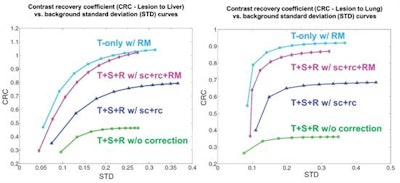 Comparison of CRC versus background STD in the reconstructed images with different levels of quantitative correction. Left: liver lesion; right: lung lesion.
To evaluate the images quantitatively, the researchers calculated the noise, measured as the standard deviation (STD) of background voxel values, and the contrast recovery coefficients (CRC) for the lung and liver lesions. Plotting CRC versus background STD showed that, for both lesions, reconstruction without any correction gave the lowest curve (poorest quality), while the two highest curves were obtained using resolution modeling. In the liver lesion, the curve for full correction and resolution modeling almost reached the CRC of the trues-only reconstruction, indicating that the applied scatter and random corrections were fairly accurate.
System comparisons
Finally, the team compared EXPLORER with an existing state-of-the-art clinical whole-body scanner. They simulated three protocols: the 36 block-ring EXPLORER with a 197 cm axial FOV; a four block-ring PET scanner with a 21.55 cm axial FOV at a single bed position; and a whole-body scan with the four block-ring system, using 33 bed positions to give a total axial FOV of 197 cm. The scan time was 20 minutes for all cases.
Of the three protocols, EXPLORER produced the best reconstructed image quality. The multibed scan produced a noisy image that did not clearly show the lesion in the liver. The single-bed image exhibited less noise, but was still noisier than the EXPLORER image.
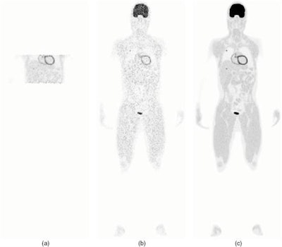 Reconstructed images from three scan protocols. (a) Four block-ring single-bed scan; (b) Four block-ring multi-bed whole-body scan; (c) the EXPLORER total-body scan.
CRC-STD curves showed that with resolution modeling, all three protocols achieved a similar maximum CRC. EXPLORER, however, provided a 6.9-fold reduction in the background standard deviation (27.9%) compared with four block-ring multibed imaging (192%), and a two-fold reduction compared with single-bed imaging (57.3%).
The researchers concluded that EXPLORER provides superior total-body PET performance at extremely low radiation doses, compared with a current clinical scanner. They are now working to further enhance their reconstruction method, by improving the image signal-to-noise ratio and reducing reconstruction time. "Other ongoing work includes development of attenuation correction methods for ultra-low dose PET and motion correction for total-body dynamic PET," Qi said.
© IOP Publishing Limited. Republished with permission from medicalphysicsweb, a community website covering fundamental research and emerging technologies in medical imaging and radiation therapy.


