FDG-PET/CT can play a valuable role in directing image-guided biopsies of pediatric cancer patients, given its ability to detect and discern between malignant and benign regions, according to a study by a group from Saudi Arabia and Canada published in the March issue of the American Journal of Roentgenology.
Among some four dozen pediatric patients with known or suspected malignancies, the researchers found 36 cases in which FDG-PET images were concordant with biopsy results, and only one case of disagreement. The findings suggest FDG-PET/CT would be useful in identifying the best site for biopsy in young patients, the group concluded (AJR, March 2017, Vol. 208:3, pp. 656-662).
"In our study, all histopathologically proven malignancies had abnormal PET results with high uptake, unless interval treatment had been administered," wrote lead author Dr. Saeed Nihayah from Prince Sultan Military Medical City in Riyadh, Saudi Arabia. "These findings support the valuable role of PET/CT in directing image-guided biopsies in children."
FDG-PET/CT's vagaries
FDG-PET/CT has long been known for its prowess in the workup of both pediatric and adult patients with suspected malignancies and/or for staging and follow-up of cancers. Positive PET/CT findings in and of themselves, however, cannot always definitively pinpoint malignancy; therefore, a biopsy may be required to determine the appropriate course of clinical management.
In addition, Nihayah and colleagues from the Hospital for Sick Children in Toronto noted that research is limited on the association between FDG-PET/CT findings and image-guided percutaneous biopsy results in pediatric patients.
Their retrospective study included pediatric patients who underwent image-guided biopsy before or after a PET/CT scan between January 2007 and December 2012. One criterion was that image-guided biopsy had to be performed within six weeks of FDG-PET/CT, either before or after the biopsy.
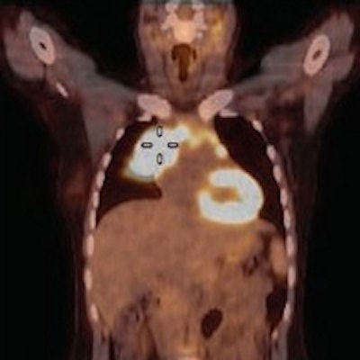
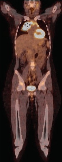
 FDG-PET/CT of a 15-year-old girl with a mediastinal mass who was a candidate for biopsy and histopathologic investigation. The coronal fused PET/CT indicates an FDG-avid tumor (cursor) in the mediastinum. Image courtesy of AJR.
Forty-five subjects were imaged on a 16-slice PET/CT scanner (Gemini GXL, Philips Healthcare) in the supine position with normal respiration. Whole-body imaging began 60 minutes after injection of 5.2 MBq/kg of body weight of FDG, up to a maximum of 370 MBq (10 mCi).
The participants were 23 boys and 22 girls, with a median age of 10.5 years (range, 4-17 years) and a median weight of 48 kg. Two patients underwent two image-guided biopsies after two FDG-PET/CT scans, so the final analysis included 47 biopsies among the 45 patients.
Two nuclear radiologists interpreted the results and were blinded to each other's evaluations. Maximum standardized uptake values (SUVmax) were obtained for each study. Any disagreements in outcome were settled by consensus among the two readers.
FDG-PET/CT-biopsy concordance
Nineteen patients (20 biopsies) were known to have a malignancy, with 12 cases of lymphoma as the most frequent diagnosis. The other 26 patients (27 biopsies) were evaluated for possible malignancy, of whom 16 were positive, including 11 patients with lymphoma. Among the 10 patients with a benign diagnosis, inflammation was the most common finding.
Among the 47 FDG-PET/CT scans, 39 (83%) showed an abnormal increase of activity at the site of biopsy. In other words, the FDG-PET result was positive, as indicated by a median SUVmax of 6.5 (range, 1.0-21.3). The remaining eight scans revealed no areas of increased activity.
Of those 39 FDG-PET/CT scans which revealed abnormal increased activity, diagnostic results were achieved in 32 cases (82%). And of those 32 diagnostic biopsies, 21 results (66%) yielded a malignant diagnosis with an average SUVmax of 6.8, compared with 11 benign diagnoses with an average SUVmax of 3.2.
In addition, among the eight PET/CT scans with no increased activity at the site of the biopsy, biopsy results were positive for malignancy in three (37%) of those eight cases.
Given the results, the researchers concluded that PET/CT can "play a valuable role" in directing image-guided biopsies for pediatric patients.
"Going forward, in future biopsies, the choice of site within a lesion or the choice of one lesion over another may be guided by results of PET, when it is available before biopsy," Nihayah and colleagues wrote.
They recommended that larger prospective studies be conducted to evaluate PET/CT's role in guiding interventional radiologists to a biopsy site and to confirm the hybrid modality's specificity and sensitivity.


