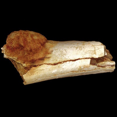
Cancer is often thought to be a product of modern times and lifestyles, but researchers have discovered what they believe is the earliest case of malignancy -- in a fossil from an early human ancestor who lived about 1.7 million years ago. The key to diagnosing this ancient cancer was advanced 3D imaging technology.
The international team of researchers used micro-CT and 3D rendering to image a hominin fossil of a metatarsal discovered in South Africa in 1948. The fossil was originally believed to have had a benign tumor, but advanced imaging techniques helped researchers re-evaluate and diagnose the bone as having malignant neoplastic disease, an osteosarcoma. They presented their findings in a case report published in the South African Journal of Science (July/August 2016, Vol. 112:7/8).
"The diagnosis has been made possible only by advances in 3D imaging methods as diagnostic aids," wrote the authors, led by Edward Odes, a doctoral candidate at the University of the Witwatersrand School of Anatomical Sciences in Johannesburg.
Rare finding
Neoplastic disease is rare in human fossils, with only a few confirmed cases dating 120,000 to 780,000 years old, according to the authors. It is also generally believed that premodern neoplastic diseases are limited to benign tumors, but this new fossil finding offers evidence that this is not the case, they noted. In their report, they presented the "earliest identifiable case of malignant neoplastic disease from an early human ancestor" that dates to 1.6 million to 1.8 million years old.
The fossil was discovered in a cave site in the Cradle of Humankind paleoanthropological site, located about 40 km from Johannesburg. The adult left fifth metatarsal specimen includes the proximal diaphysis and much of the distal section but without the articular end, according to the researchers. It has an irregular hemispherical mass that abuts the cortex and measures 5.2 mm x 4.7 mm.
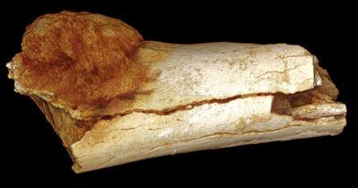 The fossil, a hominin fifth metatarsal, exhibits a hemispherical bony mass located on the proximoventral aspect of the shaft, abutting the cortical bone surface. Image courtesy of the South African Journal of Science.
Advanced imaging
Researchers originally studied the fossil and diagnosed the mass as a benign bone tumor, osteoid osteoma, but recent evaluation with advanced imaging techniques has resulted in a determination that the tumor was malignant.
The bone was scanned using a micro-CT system (XT H 225 ST, Nikon Metrology) at 100 kV and 17-µm resolution at the South African National Centre for Radiography and Tomography. Reconstruction was performed using Amira 5.4 software (FEI Visualization Sciences Group), generating 2D orthoslice and 3D surface-rendered views.
For the differential diagnosis, the researchers included various bone-forming conditions, such as chondrosarcoma, Ewing's sarcoma, metastatic carcinoma, osteochondroma, osteoblastoma, and osteosarcoma. Based on the micro-CT findings, they determined the mass to be an osteosarcoma.
Osteosarcoma is a primary malignant bone tumor that typically results in cortical and medullary disruption and some mineralization, as well as aggressive periosteal new bone reaction. Osteosarcomas tend to occur in regions of bone growth and are commonly found around the knee, but they present in metatarsals in less than 1% of modern clinical cases, the study authors explained.
The new diagnosis was supported by micro-CT imaging of a modern clinically diagnosed case of osteosarcoma of a distal femur, they noted. In comparing the images, obvious similarities were seen between the fossil and the modern case.
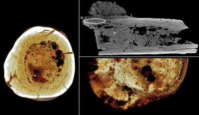 Top right: Axial micro-CT orthoslice indicating (i) reactive new bone formation subperiosteally forming a Codman triangle, (ii) ossified exophytic (cauliflower-like) and/or spiculated mass adjacent to the bone, (iii) localized subperiosteal invasion by the mass into the cortex, and (iv) remodeled bone infill. Left: Transverse rendered view of the bone. Images courtesy of the South African Journal of Science. Lower right: Transverse rendered view of a modern clinical case of osteoblastic osteosarcoma with aggressive local medullary infilling. Image courtesy of the University of Pretoria Department of Anatomy. Note the homologous combination of spongy and solid bone between the fossil and clinical specimens (v).
"This change in diagnosis is entirely the result of advances in high-resolution 3D imaging, together with the judicious use of comparative clinical pathology," the authors wrote.
More accurate diagnoses
While today's increase in cancer rates has been clearly linked with factors in the modern world such as environment, diet, and lifestyle, primary bone tumors evidently have occurred throughout history, as this case demonstrates, according to the authors.
"Modern medicine tends to assume that cancers and tumors in humans are diseases caused by modern lifestyles and environments," Odes said in a statement. "Our studies show the origins of these diseases occurred in our ancient relatives millions of years before modern industrial societies existed."
The lack of evidence of malignancies in fossil and archaeological records may be due to few samples being found, issues with preservation or analytical techniques, or inefficient imaging methods. For example, the authors noted that the results would be different if plain radiography or MRI were used instead of micro-CT.
"It is thus possible that cases of malignancy might remain unknown in fossil[s] ... awaiting imaging and discovery," they wrote. The rare number of cancer cases found in archaeological records is not representative, they argued, and should not be thought of as reflecting an accurate prevalence of the disease in ancient periods.
"Modern noninvasive imaging techniques ... play a considerable role in enabling accurate diagnoses," they concluded. "These new techniques hold considerable potential for re-evaluating previously reported paleopathological lesions."

