Breast CT with a prototype photon-counting scanner offers higher resolution than what's been reported for other types of breast imaging technologies, according to a new specimen imaging study by Willi Kalender, PhD. Another advantage of photon-counting CT is its efficient use of radiation.
In a technical feasibility study of a photon-counting breast CT system, 10 surgical lumpectomy specimens of various sizes were examined with digital mammography, digital breast tomosynthesis (DBT), and photon-counting breast CT. Additionally, a mastectomy specimen was examined to evaluate the suitability of photon-counting breast CT for larger specimens.
The research team, led by Kalender of the Institute of Medical Physics, University of Erlangen-Nürnberg, in Erlangen, Germany, found that photon-counting breast CT offers sufficiently high spatial resolution for reliable detection of calcifications and delineation of soft tissue (European Radiology, 15 June 2016).
The high resolution of photon-counting breast CT and its efficient use of radiation "is unique and a particular advantage of high-resolution photon-counting breast CT technology," the group wrote.
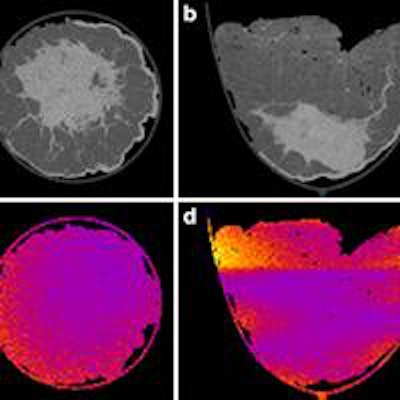
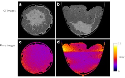
 Dose distributions are largely homogeneous, shown here for transverse and coronal views of the mastectomy specimen. All images courtesy of European Radiology.
How breast CT compares
Digital mammography is an imperfect breast cancer screening modality, especially for women with dense breasts, so researchers are always on the lookout for something new, particularly with high resolution. Kalender and colleagues aimed to investigate photon-counting CT with a novel scanner and detector concept that provides both a high spatial resolution of better than 100 µm and radiation dose levels at or below 5 mGy average glandular dose (AGD).
The researchers developed a prototype breast CT scanner that included a photon-counting detector. They then compared the unit's performance in imaging a specimen of breast tissue with results for digital mammography and DBT.
Pathological examination showed calcifications in eight of 10 lumpectomy specimens. Digital mammography, DBT, and photon-counting breast CT revealed on average 73%, 70%, and 100% of the microcalcifications, respectively.
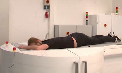
 Dedicated breast CT scanner for imaging the patient in prone position, one breast at a time.
The mastectomy specimen measured 14 cm in diameter and 9 cm across; with photon-counting CT, there were no apparent differences in image quality compared with the smaller lumpectomy specimens. Calcifications were clearly detectable in the whole volume, and soft-tissue structures were well-delineated.
Noise levels with photon-counting CT were in good agreement with laboratory simulations and were considered relatively low, which was expected because single photon-counting detectors are not very susceptible to electronic noise or other disturbing effects, the researchers wrote.
"Both noise and uniformity levels appear fully acceptable in view of the wide window settings employed in breast CT," they added. "Noise can be reduced by increasing dose in specific diagnostic applications."
Dose efficiency of the photon-counting breast CT detector was found to be very high -- supported further by the choice of an optimized x-ray spectrum that is in the range of 50 kV to 80 kV. In actual clinical use, radiation dose delivered to patients will depend on the protocol chosen and on the size of the target volume being imaged.
"The range of dose values will likely be chosen from low doses, as in screening digital mammography, similar to this study, or higher doses as in diagnostic digital mammography," Kalender and colleagues wrote. "In any case, dose distributions for photon-counting breast CT are rather homogeneous and thereby much better than in the case of projection imaging."
Specimen measurements showed excellent detection of microcalcifications for photon-counting breast CT, which clearly exceeded the results of the standard techniques of digital mammography and DBT, they added.
"This result is primarily due to the high isotropic spatial resolution offered by the photon-counting breast CT system," the authors wrote. "The general advantage of CT slice imaging over projection imaging with respect to visibility of structure details and the availability of interactive 3D reading tools routinely available for clinical CT today contribute to this."
The most favorable types of reconstruction and display still have to be determined, and further studies are planned for this year that will focus on the clinical evaluation of photon-counting breast CT, they concluded.
Study disclosures
Willi Kalender, PhD, has a relationship with CT Imaging in Erlangen, Germany.


