Nearly 10% of patients receiving a brain MRI have incidental findings, and a third of those require further evaluation for the unexpected abnormalities, according to a large Dutch study published online on June 23 in Radiology.
In the prospective study, researchers from Erasmus University Medical Center in Rotterdam found that more than 3% of some 5,800 middle-aged and elderly people in the Netherlands required substantive clinical follow-up for their unanticipated abnormalities.
Lead author Dr. Daniel Bos and colleagues also predicted an increase in the number of incidental findings in the future, since the "use of brain MR imaging in both the clinical and the research setting will continue to increase in the coming years" (Radiology, June 23, 2016).
Imaging advances
In fact, improvements in imaging technologies and greater utilization of noninvasive methods to diagnose patients have already had a profound effect on the discovery of incidental findings in many patients. If not investigated further, such abnormalities could have clinical consequences, according to the researchers.
"To date, specific guidelines on how to deal with such incidental findings on brain MR images in research settings are limited particularly because of lack of data on the magnitude and effect of the topic," the authors wrote, referencing previous studies. "Key elements needed to further the debate are information on prevalence, potential clinical relevance, and natural course of incidental findings."
During the past two decades, brain imaging has acquired a firm position in clinical practice and medical research, wrote Bos in an email to AuntMinnie.com. And although awareness of incidental findings from imaging has grown, there is still much uncertainty about how to deal with these results.
Thus, Bos and colleagues set out to estimate the frequency, severity, and outcomes of incidental findings seen on brain MRI scans.
The study included some 5,800 MRI brain exams from August 2005 through January 2014. The mean age of the study population at the first MRI scan was 64.9 years (± 10.9 years). Women comprised the majority of subjects (3,194, 55%), with a mean age of 65.1 years (± 11.1 years). The remaining 2,606 men (45%) had a mean age of 64.8 years (± 10.7 years).
All brain MRI exams were conducted on a 1.5-tesla scanner (GE Healthcare) with identical imaging protocols for all participants at each time point of the study.
Incidental findings
Among the total group of subjects, 549 (9.5%) had least one incidental brain finding. More abnormalities were found in women (306, 56%) than men (243, 44%).
The most common incidental finding was meningioma (143, 2.5%), a benign brain tumor that most often occurs in adults, followed by cerebral aneurysms (134, 2.3%). The two abnormalities accounted for more than half of all incidental findings.
The fact that meningiomas were the most common incidental finding matched previous studies, but Bos and colleagues noted that the prevalence of meningiomas was considerably higher in the current study than in previous research.
There was also a slightly higher prevalence of cerebral aneurysms in this study than in past papers, according to the authors.
"This may partly be from the use of our high-resolution proton density-weighted sequence, which permits a relatively good view of the circle of Willis compared with conventional T1-weighted and T2-weighted sequences and likely improved our detection of aneurysms," they wrote.
Other incidental findings included arachnoid cysts (92, 1.6%) and pituitary abnormalities (67, 1.2%).
Clinical follow-up
The researchers noted that 188 patients (3.2%) were referred to medical specialists for their incidental findings. For 144 patients (77%), clinicians took a "wait-and-see" approach to the abnormalities or discharged them after one follow-up exam with no need for additional care.
Fifteen meningiomas were treated invasively based on tumor growth. In addition, 16 participants with aneurysms were referred to a neurologist due to the size and location of their abnormalities; four had endovascular treatment and two had surgery.
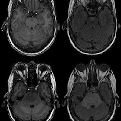
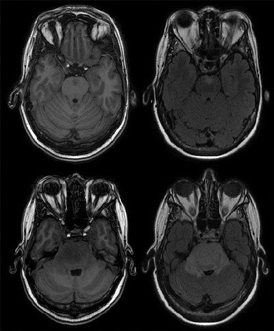 MRI brain scan shows a progressive lesion in the pons of a 50-year-old woman during two years of follow-up. T1-weighted sequence (top left) and a fluid-attenuated inversion recovery sequence (top right) find a subtle lesion in the pons. The same sequences (bottom row) of the MR exam two years later find the lesion expanded greatly. The final diagnosis was high-grade glioma of the pons. Despite radiation therapy, the patient died within 30 months of the initial examination. Images courtesy of Radiology.
Bos and colleagues were quick to mention that the prevalence of incidental findings could be even greater than they reported.
"We may have missed some small meningiomas and aneurysms because of the lack of use of intravascular contrast agent, which was not available in our study setting," they wrote. "Yet, if anything, this would mean that the true prevalence of these findings might even be slightly higher."
Disclosure debate
The issue of reporting incidental findings to patients remains an ongoing discussion, and there is no clear-cut answer to this question, Bos said. Interestingly, policies with regard to incidental findings differ considerably across different research projects in various countries.
"We believe there is no straightforward 'yes' or 'no' answer," Bos said. "The key question that should be answered first is which findings should be reported. We believe that the data we provide in the current study may serve as first basis to answer this question."
A second important question, he added, is the context in which the medical imaging is performed.
"Is the imaging part of the medical workup of a patient in the clinical setting? Or is imaging performed in volunteers taking part in medical research?" Bos asked. "These are two distinct scenarios which require differential approaches. Especially in the latter case, it is very important to acknowledge that these persons are essentially healthy."
The researchers hope these results will help guide the future management of incidental findings and provide a foundation to develop research protocols as new studies commence.
"During the future course of the Rotterdam Scan Study, new persons will be scanned and the number of persons with repeat imaging will increase. We will keep reviewing all brain MRI examinations using our standardized procedure and record any incidental findings," Bos said. "Novel insights into changes in medical regimes for these incidental findings or changes in the occurrence of specific findings will be closely monitored and may lead to further updates in the future."


