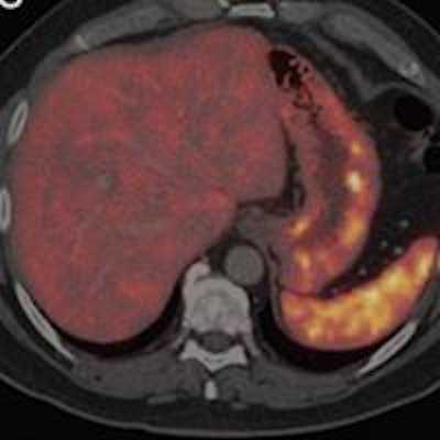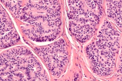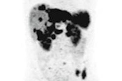
PET/CT imaging with radiopharmaceutical gallium-68 (Ga-68) DOTATATE significantly improves whole-body staging of patients with neuroendocrine tumors and should be the modality of choice in these cases, according to German researchers.
The study from the University Hospital RWTH Aachen found that 68Ga-DOTATATE-PET/contrast CT achieved greater sensitivity and specificity in the diagnostic accuracy and detection of bone metastases and lymph node metastases compared with contrast CT alone and was "very reliable" for the exclusion of suspected metastases.
Ga-68 DOTATATE-PET/contrast CT also changed the clinical management in more than 25% of patients in the study cohort (European Journal of Radiology, October 2015, Vol. 84:10, pp. 1866-1972).
Tumor recurrence
Accurate staging of neuroendocrine tumors is critical for many patients' survival, according to the researchers, led by Dr. Dirk Robert Albanus from the department of nuclear medicine at the Aachen facility. They cited an Italian study, which found that depending on the location of the tumor, survival decreases from 96% in patients with no metastases to 77% in patients with nodal, 73% in patients with liver, and 50% in patients with extrahepatic distant metastasis, such as in bone or lungs (Endocrine-Related Cancer, December 2005, Vol. 12:4, pp. 1083-1092).
The use of PET/CT with Ga-68 DOTATATE also received positive reviews last year, when a different team of German researchers confirmed the efficacy of the imaging technique for detecting the recurrence of neuroendocrine tumors.
PET/CT with Ga-68 DOTATATE achieved high sensitivity (90%) and good specificity (82%) in this clinical application. The modality also ruled out recurrence with a high negative predictive value (90%), according to lead author Dr. Alexander Haug, from Ludwig Maximilian University, and colleagues (Radiology, February 2014, Vol. 220:2, pp. 517-525).
In the EJR study, Albanus and colleagues retrospectively analyzed results from 26 men and 28 women with a median age of 64 years (range from 38 to 86 years) with histologically confirmed neuroendocrine tumors, who were available for a six-month follow-up, and had undergone a Ga-68 DOTATATE PET/contrast CT scan as part of their workup between September 2011 and March 2013.
Patients intravenously received 143 MBq (range from 92 to 185 MBq), with the Ga-68 DOTATATE PET/contrast CT scan (Gemini TF 16 PET/CT, Philips Healthcare) beginning approximately 40 minutes after injection. Images were taken with transverse and axial fields-of-views of 57.6 cm and 18 cm, respectively.
Researchers considered a PET-positive lesion as an area with greater intensity than the background and registered a maximum standardized uptake value (SUVmax) of greater than 2.7 for bones, 2.2 for lymph nodes, and 2.3 for pulmonary lesions. The values were chosen because they represent the upper limit of physiological uptake in these regions.
To evaluate results, two nuclear medicine specialists and two radiologists initially analyzed PET and contrast CT images separately. All four readers then performed a consensus reading using a five-point grading system in which positive lesions were deemed grade 3 or 4, and negative lesions were grade 2 or less.
Researchers then used clinical and follow-up data (median of 12.6 months, range from 6.1 to 23.2 months) as the reference standard to compare contrast CT and the PET/contrast CT results and classify lesions as true-positive, false-positive, true-negative, or false-negative. Follow-up included a repeat PET/CT or CT, an MRI, and clinical evaluation to measure the tumor.
Lesion analysis
In the tally of bone lesions, PET/contrast CT detected 139 true-positive bone lesions, compared with 48 true-positive bone lesions by contrast CT alone. The lesions were found among 17 patients identified as positive in follow-up. There were no false-negative patients with PET/contrast CT. Among those 17 patients, contrast CT alone classified eight patients as true-positive and nine patients as false-negative.
| Bone lesion detection | ||
| Ga-68 DOTATATE PET/contrast CT | Contrast CT | |
| Sensitivity | 100% | 47% |
| Specificity | 89% | 49% |
| Positive predictive value | 81% | 30% |
| Negative predictive value | 100% | 67% |
PET/contrast CT also discovered 106 true-positive lymph node metastases, compared with 71 detections for contrast CT alone. The metastases were found by PET/contrast CT in 23 patients, who all were identified as positive in follow-up, leaving two patients as false-negative. Contrast-enhanced CT alone found at least one true-positive lesion in 16 patients, with nine patients deemed as false-negative. Of 29 true-negative patients, PET/contrast CT showed at least one positive lymph node in five patients, compared with 12 positive cases for contrast CT alone.
| Lymph node detection | ||
| Ga-68 DOTATATE PET/contrast CT | Contrast CT | |
| Sensitivity | 92% | 64% |
| Specificity | 83% | 59% |
| Positive predictive value | 82% | 57% |
| Negative predictive value | 92% | 65% |
In the lung evaluation, PET/contrast CT and contrast CT both detected at least one metastasis in 10 patients, who proved to be positive during follow-up. Neither modality missed any lesions. PET/contrast CT and contrast CT alone both detected all 26 lung metastases.
| Lung lesions detection | ||
| Ga-68 DOTATATE PET/contrast CT | Contrast CT | |
| Sensitivity | 100% | 95% |
| Specificity | 100% | 82% |
| Positive predictive value | 83% | 56% |
| Negative predictive value | 100% | 100% |
Patient management
The findings of positive lesions in seven of nine patients with additional bone metastases resulted in therapy not previously planned.
PET/contrast CT showed at least one lymph node metastasis in seven patients, who were deemed negative with contrast CT alone, with two patients undergoing therapy. Two other people were operated on again with resection of lymph node metastases. The disease was upstaged for three individuals through PET/contrast CT with no further direct clinical consequences.
Based on the results, Albanus and colleagues concluded that Ga-68 DOTATATE PET/contrast CT "significantly improves" whole-body staging of patients with neuroendocrine tumors. "Therefore, Ga-68 DOTATATE PET/contrast CT should be the imaging modality of choice in patients with neuroendocrine tumors."


