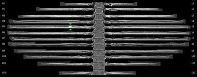
An automated curved planar reformatting software tool that presents the ribs in an "unfolded" view is starting to prove its clinical value, according to a presentation at the European Society of Thoracic Imaging (ESTI) annual meeting, held last month in Amsterdam.
"Rib-unfolded views can potentially improve efficiency in evaluating the ribs, but familiarization with normal appearances and an understanding of its limitations are necessary," noted Dr. Blake Niederhauser and colleagues from the department of radiology at Mayo Clinic in Rochester, Minnesota, U.S. "The software, however, is not perfect, and numbering errors and reformatting distortions such as rib combining and partial rib exclusion occur at low rates.
 Curved planar reformatted or "unfolded" rib view image processed with the syngo.CT Bone Reading application. Image courtesy of Siemens Healthcare.
For clinical use, they recommend bearing in mind the following key points:
- Initial scan of axial images should be performed to identify cervicothoracic anatomic variations, supernumerary, and bifid ribs, and to ensure proper automated segmentation.
- The costochondral junctions and rib segments medial to the costotransverse junction should be scrutinized primarily with axial views.
- Lateral fractures and any fracture with any degree of displacement are readily identified with unfolded views.
- Rib unfolding in patients with motion artifact or scoliosis often is misregistered.
In an e-poster at ESTI 2014, they sought to highlight the advantages and pitfalls that the commercially available reformatting tool from Siemens Healthcare, syngo.CT Bone Reading, offers in evaluating rib lesions. They also provided tips for incorporating unfolded views into clinical practice.
Because of their multiplicity and complex shape, following and numbering ribs using conventional planar reformatting is tedious and time-consuming, according to the authors. As a result, rib lesions, including fractures and metastases, are sometimes misnumbered in reports or altogether missed in up to 20% of instances, and such errors can result in improper oncologic staging or operative repair. 3D reformats provide a good overall view, but generally are not useful for primarily identifying subtle fractures or metastatic lesions.
"Rib-unfolding software provides automated numbering on both unfolded and conventional multiplanar reformatted views and provides cross-linking between the two for troubleshooting," they stated. "In unfolded views, two options for scrolling are available: a 'propeller' or 'angular' mode, which spins the ribs reminiscent of horizontal window shades, and an 'in-plane' or 'volume' mode, which allows 'in-and-out' scrolling through the rib volume."
On a standard computing platform, automated reformats take about a minute to perform in the background and can be used for rapid identification and reporting of rib lesions. To track each rib individually using standard axial views, at least 24 "up-and-down" scroll patterns are required. When a lesion is detected, particularly when attempting to view multiple ribs simultaneously, the Mayo Clinic group finds it is common to forget the rib number, and it requires additional time to recount the ribs to the abnormality.
Rib-unfolded views and automated rib numbering provided with this tool can simplify and accelerate interpretation and reporting of rib lesions, including fractures and metastases, they explained. All the ribs can be evaluated simultaneously at a glance, and areas of question in multiple segments can be evaluated with a single scroll and cross-referenced to the corresponding standard multiplanar images.
"When multiple lesions, such as rib fractures or metastases, are present, unfolded views provide rapid identification and accurate numbering. When suspicion for fractures is low, a negative exam can be quickly identified," they wrote. "Many users in our research practice commented that the numbers which track with ribs while scrolling through axial images was perhaps as useful as unfolded views given that cross-referencing to axial views is often required anyway."
When evaluating ribs for metastatic disease, they advise caution in overcalling normal rib heterogeneity for malignancy. With the new mode of rib viewing, radiologists need to have enough experience with normal variation to avoid this pitfall.
Problems and pitfalls for clinical use
In general, unfolded views provide an accurate representation of the ribs, and rib lesions are quickly and accurately identified, the authors noted. But they have identified several problem areas that can result in false-negative (missed lesion detection) and false-positive (artifact) results:
Scoliosis or minor thoracic curvature: For instance, standard coronal (upper) and rib unfolded (lower) views of a patient with acute chest trauma demonstrating moderate thoracolumbar scoliosis can result in misregistration and truncation of ribs on the right on unfolded views. Such errors can be corrected manually, but when multiple errors are present, the process can be tedious and time-consuming.
Supernumerary, variant, and bifid ribs: A 3D-reformatted (upper) view can shows 13 rib pairs, including variant pseudoarticulation of the right upper rib with the second right rib, but these anomalies may be ignored by rib-tracking algorithms as unfolded views (lower) are seemingly normal and display only 12 pairs. In patients with these types of anatomic variation, clear descriptions of the anatomy and manual counting of ribs are necessary as unfolded views may be misleading. Manual addition of supernumary ribs can be performed but may be time-consuming.
Anterior fractures, subtle buckle fractures, and fracture of the costochondral junctions: A good understanding of the appearance of these subtle fractures is necessary for accurate interpretation.
Posterior fractures: In a patient with acute thoracic trauma, identifying the trauma on unfolded views can be difficult because of reconstructive distortions, rib abnormalities posteromedial to the costovertebral junction.
Beam-hardening artifact: A dense object lying external to a patient's right chest may create streak artifact through the right eighth rib. On corresponding unfolded rib views, this results in the false-positive appearance of callous formation suggestive of a healing rib fracture.
Motion artifact: In a patient with acute chest trauma and multiple right-sided rib fractures, respiratory motion artifact in the upper thorax results in misregistration and truncation of multiple upper ribs bilaterally.
Nondisplaced acute rib fractures, while possibly less clinically relevant, are often only evident on unfolded views in retrospect, and detection remains problematic as in traditional viewing methods, the authors concluded. Normal ribs contain significant heterogeneity and a high incidence of benign lucencies and scleroses, and users should become familiar with normal variation prior to evaluation for metastases.


