The utility of optoacoustic or photoacoustic imaging in preclinical research has been established and researchers are now working toward clinical translation of the modality. However, 2D scanners typically use linear arrays that suffer from poor resolution in the direction perpendicular to the scan plane, and this limits the diagnostic power of 3D images reconstructed from 2D data acquired along that axis.
In a new study, researchers in Germany have tackled this issue with a bidirectional scanning approach that improves the elevational resolution of 3D images by acquiring data along two perpendicular axes, instead of the single axis conventionally used (Journal of Biophotonics, 15 April 2014).
"Our motivation was to improve on this issue and provide images with isotropic resolution," said senior author Vasilis Ntziachristos of Technical University Munich (TUM), who carried out the study with postdoctoral student Mathias Schwarz and collaborator Andreas Buehler, who are based at Helmholtz Centre Munich.
Taking a closer look
The researchers envisage the bidirectional technique will be used as an adjunct to conventional optoacoustic scans, which are conducted with a handheld probe. With a higher frame rate, the conventional scan allows a large volume of tissue to be examined quickly in real-time, while the higher resolution bidirectional approach enables more detailed examination of volumes of interest.
 Mathias Schwarz and Andreas Buehler from Helmholtz Centre Munich examine the imaging results.
As in conventional optoacoustic imaging, the bidirectional technique scans the linear array perpendicular to its 2D scan plane in one direction. The array is then rotated 90° to perform a second scan of the same volume. Each scan is reconstructed using filtered back projection and a composite 3D image is generated by taking the square root of the voxel-by-voxel multiplication of the two. This approach suppresses noise and arc artefacts -- a byproduct of back projection -- that occur in the individual scan sets.
Using experiments and simulations, the researchers quantified the improvements in image resolution provided by the technique over conventional unidirectional scans. Both investigations used phantoms containing microspheres ranging in diameter from 10 mm to 80 mm, and a further physical phantom containing sutures, to assess the effect of the scanning technique on line-like objects.
The researchers used a 128-element linear array focused in the elevational direction at a depth of 7.5 mm by an acoustic lens. It was scanned in 30-mm steps across an 8 x 8-mm area, using translation and rotation platforms in the experimental study. A pulsed optical laser coupled to two fiber-optic bundles was used to illuminate the phantoms.
Acquisition of the two 3D data scans took around a minute and image reconstruction took approximately five to eight minutes. A calibration corrected for deviations from the ideal geometry for the pair -- where the transducer's main axis is perpendicular to its direction of travel and parallel to the horizontal -- to prevent image distortion.
Large gains in resolution
The simulations and experiments produced similar results: A significant gain in elevational resolution was obtained using the bidirectional approach. For example, in the experimental study, the bidirectional approach resulted in a full width at half maximum (FWHM) of 107 mm, compared with 282 mm for the conventional scan. A small deterioration in the lateral resolution of the bidirectional images from a FWHM of 76 mm to 104 mm, a byproduct of the image reconstruction approach, also was seen.
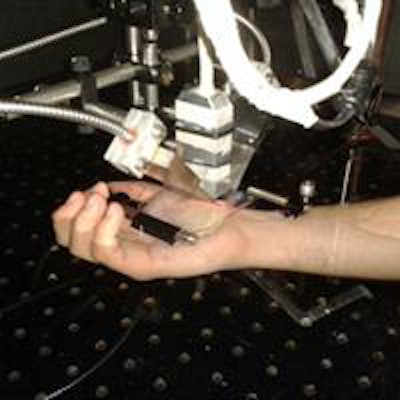
 Bidirectional optoacoustic imaging of a volunteer's hand using the scanning prototype.
Improvement in image quality was also clear when the researchers inspected the bidirectional images visually. The microspheres were correctly reproduced as points, unlike in the conventional scan where they were significantly elongated in the elevational direction. The superior resolution of the bidirectional approach was also visible in images of a chicken breast with a 4-mm deep burn.
Clinical applications of optoacoustic imaging, which can penetrate significantly deeper than purely optical techniques such as optical coherence tomography, include breast and vascular imaging. However, the major focus of the TUM group is dermatology. "We expect the [bidirectional] method to yield improved visualization of burns, wounds, and probably the invasion of melanoma," lead author Schwarz said.
© IOP Publishing Limited. Republished with permission from medicalphysicsweb, a community website covering fundamental research and emerging technologies in medical imaging and radiation therapy.


