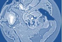
In screening for breast cancer, conventional mammography carries a finite risk of false positives and undetected lesions, motivating researchers to look for more accurate imaging approaches. Phase-contrast CT is a promising candidate that exploits variations in x-ray phase, instead of absorption, to produce superior soft-tissue contrast to conventional CT imaging.
In new work, researchers in Germany and France have imaged samples containing healthy breast tissue and four tumors, documenting phase-contrast Hounsfield unit (HUp) values for each tissue type. Based on quantitative analysis alone, healthy tissue was distinguishable from malignant, but malignant tumors were indistinguishable from benign lesions. There were, however, variations in tumor morphology and the researchers concluded that combined assessments of HUp and morphology using the modality hold the most clinical promise (Physics in Medicine and Biology, 10 March 2014, Vol. 59:7, pp. 1557-1571).
First author Marian Willner from the Technical University of Munich (TUM) and co-authors used x-ray grating interferometry, which has two key advantages over other phase-contrast approaches. "It works efficiently with standard laboratory x-ray tubes with a polychromatic spectrum and provides quantitative values for electron densities, which proves more challenging with other phase-contrast methods," said co-author Julia Herzen, who is also based at TUM.
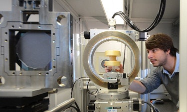 First author Marian Willner from TUM.
The researchers imaged four tissue samples, excised from patients undergoing surgery to remove tumors. The samples included healthy adipose and fibroglandular tissue and variously contained benign phylloides and fibroadenoma tumors and malignant lobular and ductal carcinomas.
Three of the samples were scanned using a standard polychromatic x-ray tube in a laboratory at TUM. The fourth, containing the invasive ductal carcinoma, was imaged with a monochromatic 23 keV beam using the ID19 beamline at the European Synchrotron Radiation Facility in Grenoble, France.
','dvPres', 'clsTopBtn', 'true' );" >
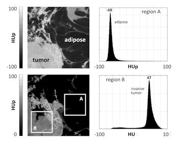
Click image to enlarge.
In 15 x 15 mm² images acquired using a synchrotron, phase-contrast CT shows good contrast between adipose tissue (peaking at -68 HUp) and invasive ductal carcinoma (peaking at 47 HUp). Bright dilated ducts are also visible in the tumor. The marked boxes measure 5 x 5 mm².
In each case, the HUp values were derived from tomographic images reconstructed from projections acquired over 360° by rotating the tissue sample. The high-resolution synchrotron images were used to measure reference HUp values for the two healthy tissues and the ductal carcinoma, enabling comparison with laboratory-based measurements and HUp values derived from data in the literature. Histological analysis identified the different tissue types in the samples.
Characterizing tissue types
Averaging over 24 10 x 10 pixel regions-of-interest for each tissue type, distinct HUp values of -68.7 (standard deviation 2.7), 65.2 (standard deviation 6.0), and 47.0 (standard deviation 2.8) were measured for adipose tissue, fibroglandular tissue, and ductal carcinoma, respectively. Higher values of 72.5 (standard deviation 5) were observed in subvolumes of the tumor that contained dilated ducts, seen as bright circular regions in the images.
Images of the fibroadenoma acquired using the simpler laboratory-based set-up showed another tumor-specific morphological feature: bright fibrous strands around 20 HUp higher than the surrounding tumor.
HUp values derived from the laboratory set-up were consistent with the synchrotron data and in all tumors, peaked between 45 and 50 HUp. However, while values for adipose tissue generally agreed with values derived from data in the literature, significant differences were seen for fibroglandular tissue.
','dvPres', 'clsTopBtn', 'true' );" >
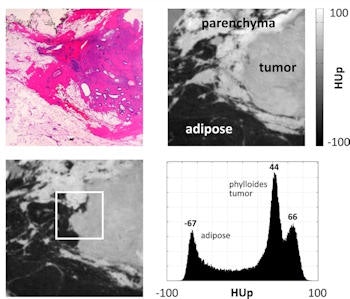
Click image to enlarge.
Phase-contrast images acquired with a standard x-ray tube in the lab (top right and bottom left) contained HUp values that matched those measured at the synchrotron. Histological analysis was used to identify the different tissue types.
"These are most likely due to the higher spatial resolution of our method revealing more morphological details, which are averaged using other methods," Herzen said.
Herzen told medicalphysicsweb the HUp values they measured define the electron density resolution needed in tomorrow's clinical scanner. "It should be able to resolve 15-20 HUp to distinguish between different tissue and tumor types." Fed into simulations, the measured tissue characteristics can also improve the accuracy of patient dose predictions for x-ray breast imaging techniques in general.
In ongoing research, the group is examining HUp values and tumor morphology in a larger patient sample, as well as trialing advanced acquisition techniques and iterative reconstruction algorithms to increase imaging speed and reduce patient dose, respectively. The researchers are also using their measured data to inform the design of phantoms for further testing of phase-contrast CT and phase-contrast mammography, which is the closer of the two to clinical implementation.
© IOP Publishing Limited. Republished with permission from medicalphysicsweb, a community website covering fundamental research and emerging technologies in medical imaging and radiation therapy.


