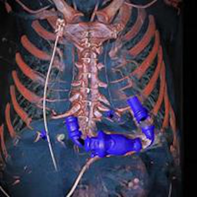
A Swedish researcher has received the top prize in a prestigious image competition for his 3D dual-energy CT (DE-CT) scan of a patient who received a mechanical heart pump while waiting for a heart transplant.
Dr. Anders Persson was named the overall winner in the 2014 Wellcome Image Awards competition, which recognizes "the most informative, striking, and technically excellent images" recently acquired by Wellcome Images, a U.K.-based resource for medical imagery. Persson, a radiologist, is director of the Center for Medical Image Science and Visualization and is a professor at Linköping University in Sweden.
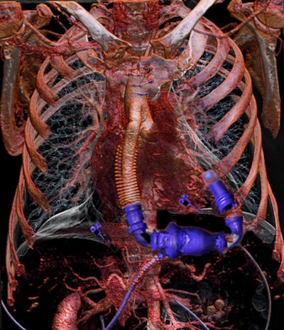 A 60-year-old man was examined with dual-energy CT (Somatom Definition Flash, Siemens Healthcare) in 2012 at the Center for Medical Image Science and Visualization (CMIV) at Linköping University. Dual energy was used to reduce metal artifacts from the pump. A new way to enhance the image quality and reduce noise was then added, and raw data from the CT were processed with iterative reconstruction software. The resulting image dataset was finally produced using volume-rendering 3D. This high-resolution image, similar to the version Wellcome awarded, won an award from the Royal Photographic Society and was selected for the International Images for Science Exhibition 2013. Image courtesy of Dr. Anders Persson.
The image that won the award was a 3D reconstruction of a DE-CT scan, with the heart pump, breastbone, collarbone, and bones of the rib cage made opaque. Other layers of tissue were made see-through so the connections between the heart and the pump can be checked.
 Dr. Anders Persson.
The heart pump is wired to the left side of the diseased heart and to the aorta, and the image reveals that the pump's connection to the heart is "faultless," according to Wellcome's description of the image. The DE-CT technique is useful both for noninvasively diagnosing medical conditions as well as for performing virtual autopsies.
Persson's image was chosen for both its technical detail as well as what it revealed of the human anatomy, according to Fergus Walsh, medical correspondent for the BBC and one of the Wellcome judges.
"It was very hard work picking an overall winner, but we were especially impressed by Anders Persson's 3D image of a mechanical heart fitted inside a human chest," Walsh said. "The juxtaposition of delicate human anatomy with the robust mechanical plumbing parts was striking, and the image was rendered so vividly in 3D that it appears to jump out at the viewer."
Other notable images in the awards included a scanning electron micrograph of a zebrafish embryo, a light micrograph of a lily flower bud, a CT scan of a seal head, and a micro-CT scan of a medieval human jawbone.


