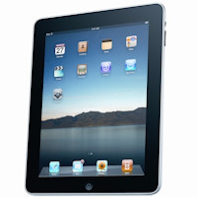
Apple's third-generation iPad can perform similarly to a 3D PACS workstation for the task of interpreting emergency CT studies, according to researchers from Hannover Medical School in Germany.
In research published online 26 July in RöFo, a team led by Dr. Susanne Tewes found no statistically significant difference in interreader variability for interpreting two typical emergency CT studies.
"From a clinical standpoint, there are potentially attractive application areas for the [third-generation iPad] primarily with respect to quick preliminary evaluation on duty or for real-time consultation of a radiological background service or experts," the authors wrote.
Due to regulatory conditions in place in Germany, the iPad can't currently be used for image interpretation in that country. However, since the third-generation iPad's high-resolution display largely meets the requirements of the current quality assurance guidelines in the German X-Ray Ordinance and DIN Prestandard 6868 -- 57 standard, the researchers sought to determine if the new-model iPad could be usable for detecting two typical issues encountered in radiological emergency diagnosis.
CCT and CTPA studies
For their retrospective study, the researchers gathered 20 native cerebral CT scans (CCT) that were acquired between October 2010 and May 2012 for evaluation of early signs of infarction. The studies were acquired during the night or weekend shift and showed one or more early signs of infarction, such as hyperdense middle cerebral artery sign, loss of insular cortex, inability to differentiate between gray and white matter, and sulcus effacement. The team also selected 20 additional native cerebral CT scans with no abnormal acute ischemic findings for a total of 40 images.
All CT scans were acquired on 16-detector-row CT scanners using a sequential scan technique and orbitomeatal angulation of the gantry. Two datasets with a slice thickness of 2.5 mm and 5.0 mm were reconstructed from the raw data using filtered back projection, according to the researchers.
The team also selected 20 CT pulmonary angiograms (CTPA) for evaluation of pulmonary embolism that were acquired from May 2011 to May 2012 during the night or weekend shift. These cases displayed acute pulmonary artery embolisms on the segment or subsegment level. As with the CCT scans, 20 CTPA studies without evidence of embolism also were included.
All the CTPA exams were produced on a VCT 64-detector-row scanner (GE Healthcare) with injection of Iomeron 350 contrast (Bracco Diagnostics). Image data were reconstructed with a slice thickness of 1.25 mm and a reconstruction interval of 1.0 mm via a standard filtered back projection algorithm, the researchers said.
Three radiologists were then asked to read the exams, first on a Z800 standard PACS workstation (Hewlett-Packard) with a calibrated RadiForce RS210 display (Eizo Nanao Technologies) running Visage 7.1 3D thin-client software (Visage Imaging). The radiologists had three, four, and six years of experience, respectively, in reading emergency CT scans.
With a reading interval of at least three weeks later, the radiologists again read the rerandomized datasets on a third-generation iPad running the Visage Ease mobile image display application (Visage).
In both sessions, the readers evaluated the image data for either the presence of early signs of infarction or a pulmonary artery embolism using a five-point Likert scale ranging from 5 (definitely pathological) to 1 (definitely not pathological). The readers were blinded to both the clinical findings and the relative proportion of scans with pathology. They did know, however, that the frequency distribution did not correspond to the clinical routine.
For all three readers, there was no significant difference in inter-reader variability for either the cerebral CT or CTPA scans (p > 0.05).
In agreement with several prior studies, the authors said they consider a potential role for the third-generation iPad in radiology primarily as a supplement to established workstations.
"Mobile access to image data allows quick preliminary initial assessment as well as teleconsultation (e.g., of the radiological background service) under emergency conditions," the authors wrote.
Not in Germany
Despite these findings, don't expect the latest generation of iPad to be used clinically for viewing images in Germany anytime soon. While the U.S. Food and Drug Administration (FDA) has already cleared multiple iPad applications for diagnostic review of images if a PACS workstation is unavailable, similar approvals can't be expected in Germany, according to the researchers.
"Although the [third-generation] iPad fulfills significant parts of the currently valid quality assurance guidelines, it does not reach the required minimum diagonal screen size of 15" (LCD)," the authors wrote. "The inconstant environmental conditions associated with mobile operation are also problematic even though similar concerns on the part of the FDA were able to be resolved by software solutions for ensuring appropriate contrast detectability."
Requirements for approval and constancy testing of image display devices are also expected to increase further with the upcoming amendment of the quality assurance guidelines and pending new standard DIN 6868 -- 157, according to the researchers.
Other challenges to the use of mobile devices such as the iPad in radiology include ensuring appropriate background lighting and also appropriate data security. Users would also need to guard against screen contamination due to its use of a touchscreen, according to the researchers.
"Possible application areas are currently teaching, demonstration of diagnosed images to colleagues or patients, or use as a medical information system and expert system," the authors wrote.


