Philips Healthcare's Achieva TX MRI system and its Avalon fetal monitor will be used at sites in the U.K. to map fetal brains and babies' brains after birth.
Scientists from Guy's and St. Thomas' hospitals, King's College London, Imperial College London, and the University of Oxford are participating in the project to investigate the wombs of pregnant women and image their babies' brains, even while the fetuses are moving.
The researchers will map how the human brain grows and forms connections with the aim of mapping for the first time how the brain assembles itself, and also observe how brain connections and patterns of activity seen on older subjects emerge as babies grow in the womb and just after birth. Up to 1,500 subjects will be studied to both examine normal development and explore early signs of disabilities, such as autism and attention deficit disorders.
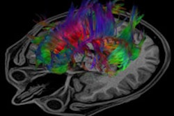
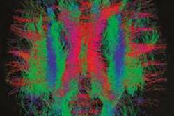
The objective is to create a "connectome," a kind of wiring diagram of the fetal and infant brain that shows the formation of brain structures, such as the cerebral cortex or the hippocampus, and the connections between them, Philips said.
To enable the project, new MRI facilities have been installed at St. Thomas' Hospital, creating a dedicated imaging suite, integrated with the neonatal critical care unit and a wide-bore scanner for fetal imaging.


