Trimodality PET/CT-MR imaging using a novel system to shuttle patients between scanners offers new insights into the diagnosis of cancer patients. How the configuration is being used at the University Hospital Zurich is described in an 8 October article in Magnetic Resonance Materials in Physics, Biology, and Medicine.
The primary advantages of being able to transfer a patient directly from an MRI modality to a PET/CT modality have clinical, cost, and workflow efficiency benefits, which the university's department of medical imaging has been experiencing since November 2010.
 Lead author Dr. Patrick Veit-Haibach.
Cost is one compelling feature. The total cost of purchasing separate MRI and PET/CT scanners, with a dedicated bed to shuttle patients between them, is estimated be between 3 million euros ($4 million U.S.) and 5 million euros ($6.5 million), whereas the cost for an integrated PET/MRI system located in a single room is approximately 5 million euros just by itself.
Scheduling and utilization of modalities are also very important to a medical imaging department. The MRI and PET/CT scanners used in the trimodality configuration can be scheduled independently at the same time for optimum utilization. Having an MRI exam performed during the uptake period of the PET tracer prior to the PET/CT exam is also more efficient for the patient.
The department takes advantage of this flexibility. It schedules five to six patients in the morning for trimodality imaging in 60-minute time periods starting at 8 a.m.
"We start with neuro scans or joints at 7:45, so we have one or two scans already done before the first PET/CT-MRI patients get to the MRI suite. We then perform the MRI during the FDG uptake time. In the afternoon, we use each scanner for individual exams, allocating 40-minute time slots," according to lead author Dr. Patrick Veit-Haibach.
In a clinical trial testing the technique, approximately 80% to 90% of patients who were prescribed a PET/CT had the examination as a PET/CT-MRI study in September. Veit-Haibach expects that approximately 75% of patients having a PET/CT -- or between 70 to 75 patients -- will have the trimodality exams performed. Two other PET/CT scanners also are in use in the department.
Dedicated patient transfer shuttle and patient positioning
Veit-Haibach, a dual board-certified radiologist and nuclear medicine physician who heads the PET/CT-MRI section of the department of medical imaging, attributes the success of trimodality imaging to having a dedicated patient shuttle system to enable accurate multimodal image registration. They currently use a custom-built side-loading shuttle (Innovation Design Center, Thalwil, Switzerland) that consists of a metal trolley and fiberglass transport board. The board carrying the patient is held by two supporting sliding arms and can flexibly be slid either to the left or right.
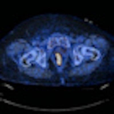
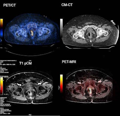
 Left-sided, FDG-avid rectal cancer with rectal wall thickening. Note the improved detectability of the surrounding infiltration and detectability of the small locoregional lymph node. Images courtesy of Dr. Veit-Haibach.
After the board is positioned over the scanner table, the table is elevated until it supports the board. The board is released from the sliding arms and the images are acquired. Upon completion of the exam, the shuttle is positioned next to the scanner table and the arms are slid underneath the transport board. When the scanner table is lowered, the patient is transferred back onto the shuttle.
This process has been made a bit easier with the replacement of a standard-bore MRI system (Discovery MR750 3-tesla, GE Healthcare) with a wide-bore magnet (Discovery MR750w 3-tesla). This was done in February 2012. The same PET/CT system (Discovery PET/CT 690, GE Healthcare) has been used since November 2010.
"We always coregister the PET/CT images with the MRI data using a commercially available imaging workstation (Advantage Windows, GE Healthcare). The MRI exam is typically performed as a whole-body examination due to current research trials comparing PET/CT and PET-MRI. It also may be a partial-body contrast-enhanced examination of the abdomen, pelvis, head/neck, or brain," Veit-Haibach said.
The trimodality PET/CT-MRI configuration can use standard CT-based PET attenuation correction. This is an important advantage in terms of accurate and robust PET quantification. The sequential approach allows selective use of radiofrequency (RF) coils during MR scanning, as well as their removal during PET/CT without moving the patient.
The authors consider this an important advantage because RF head coil structures can cause significant attenuation artifacts. The authors also recommend that posterior coils are integrated into the MRI system table to avoid patient repositioning during the selective placement or removal of the RF coils.
Clinical value
Trimodality imaging offers clinical advantages when imaging lymphoma, myeloma, and melanoma due to its combined ability to identify more cancers and see them more clearly. When following patients after treatment for multiple myeloma, for example, PET/CT has advantages in detecting cortical lesions, but whole-body MRI can visualize lytic bone marrow lesions more effectively.
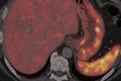
While both PET/CT and MRI can detect liver lesions, the authors have found that lesion conspicuity is better seen in MR images. Also, the diagnosis of neuroendocrine tumors of the gastrointestinal tract may be improved with trimodality imaging, as well as making a more accurate diagnosis of active versus inactive inflammatory lesions, the authors wrote.
For prostate imaging, the combination of PET/CT-MRI can serve as a one-stop imaging tool for patients who need staging and restaging. The images provide information about the local tumor status, nodal status, and distant metastases.
Patients with head and neck cancers also benefit. MR images show the infiltration of tumors into surrounding tissues, and PET/CT can detect lymph node metastases better.
"Trimodality imaging also has advantages in patients who exhibit metal artifacts in CT, as new MR sequences can minimize those artifacts. We believe for this reason head and neck cancer is one of the most promising indications for a PET/CT-MRI or simultaneous PET/MRI," Veit-Haibach said.
Trimodality imaging may be useful in clinical trials, the authors added. Patients may be easier to acquire for comparison trials of PET/CT and PET/MRI in a clinical routine setting because a patient may be transferred using a dedicated shuttle system directly to the PET/CT following the first exam with no differences in SUV.
The authors' primary goal in clinical neuroimaging is to combine quantitative PET and MR imaging and to establish this multimodality procedure in routine imaging of brain pathology. How will this be coordinated?
Veit-Haibach told AuntMinnieEurope.com that unlike hospital departments with separate sections for MRI and PET/CT, University Hospital Zurich has established a section in the department of medical imaging exclusively for trimodality imaging. The resident training program in this subspecialty takes eight years to complete. Veit-Haibach also oversees radiologists who, while not specializing in nuclear medicine, are working on multimodality imaging projects.
Trimodality imaging programs are just starting to be implemented. Veit-Haibach is aware of systems in China, Denmark, the Middle East, and South Korea. "Interest is increasing," he said. "We host visitors from around the world on a regular basis."


