As radiotherapy delivery techniques continue to advance and new systems emerge, have we reached the point at which it's possible to accurately deliver the planned dose distribution? At last month's annual meeting of the European Society for Radiotherapy and Oncology (ESTRO) meeting in Barcelona, experts addressed this question.
At the dedicated symposium titled "Can we deliver the dose distribution we plan?" three speakers examined the current limitations on defining and delivering required dose distributions, with regard to their respective areas of expertise: external-beam x-ray radiotherapy, ion beam therapy, and brachytherapy.
The discussion kicked off with a presentation from Dr. David Thwaites, director of the Institute of Medical Physics at the University of Sydney in Australia. He examined the situation for megavoltage x-ray radiotherapy. He pointed out that while sophisticated modern technologies are able to deliver almost anything, there remains a host of unanswered questions.
 Session speakers Dimos Baltas, Martijn Engelsman, and David Thwaites.Image courtesy of Tami Freeman.
Firstly, do we actually know the appropriate dose distribution to deliver to an individual patient? "Currently," said Thwaites, "No, we don't, as we don't understand the radiobiology well enough on an individual patient basis."
Multimodal and functional imaging can help, but limitations remain due to fusion uncertainties and less than optimal image quality. As such, much research effort is currently going into improving deformable image registration techniques.
Inconsistent target delineation between observers can also cause difficulties, as can the fact that modern delivery techniques, such as intensity-modulated radiotherapy (IMRT), volumetric-modulated radiotherapy, and image-guided radiotherapy, alter the dose-volume trade-offs, the constraints and the margins. For example, IMRT produces a different dose spread to previous delivery techniques, with larger low-dose regions, the implications of which are not yet fully understood.
The next question asked if we can model the required dose distribution accurately and identify the best treatment plan. "Just about," said Thwaites. "In terms of plan optimization, we need to balance quality versus delivery limitations and delivery efficiency," he explained. Other factors to consider include: whether to perform biological or physical optimization; whether uncertainty can be included in the planning process; and which delivery technique and plan is best for a particular case, using multicriteria approaches. Accurate low-dose modeling remains a problem.
And once we know what dose to deliver, can we realistically and robustly deliver it? "Yes, we can," Thwaites said, adding the caveat that it's vital to use image-guidance for complex treatments and to verify dose delivery.
At present, most radiotherapy is performed using pretreatment verification, but in vivo dosimetry could detect errors that may otherwise be missed. "We should be doing in vivo dosimetry with electronic portal imaging devices," he explained. "This is not fully supported by manufacturers at the moment, but clearly, it's where we should be going."
Another issue that must be addressed is the ability to deal with inter- and intrafractional variations. Adaptive radiotherapy is still very much in development, and can be dependent upon the DIR algorithm chosen. For larger motion changes, tracking systems are reported to work reasonably well, but are not yet at the point of widespread implementation. There is also a need for more efficient and integrated quality assurance tools and methods.
"Yes, we can deliver the dose distribution that we plan," Thwaites concluded. "But there are still significant uncertainties and significant research challenges. What's more, different developmental steps are interdependent -- as one improves, it affects the others."
Proton observations
The next speaker to take the podium was Martijn Engelsman, PhD, a medical physicist from the Holland Particle Therapy Centre in Delft, the Netherlands. Engelsman addressed some of the issues surrounding proton and ion-beam therapy. While protons offer highly conformal dose delivery, he explained, the planned dose distribution is much more sensitive to set-up errors and anatomy variation.
Proton therapy is currently delivered using either pencil-beam scanning or passive scattering. Of the two, scanning offers higher target conformation, but is affected more by intrafractional target motion. Intensity-modulated proton therapy (IMPT) offers the best conformity, but is most sensitive to anatomy variation.
"Delivering dose to a phantom works well, the machine is not the problem, you can deliver what you want," he explained. "But things become different in a patient." Clinical treatments introduce the need to deal with set-up errors, range uncertainties, and density changes that impact the beam range. For pencil-beam scanning, interplay effects between the patient's breathing motion and the beam motion also impact the delivery accuracy.
Engelsman described some of the countermeasures that are available for dealing with motion and interplay. For range uncertainties and set-up errors, options include robust optimization of treatment plans and, to a lesser extent, the use of safety margins. The move to robust optimization is technically solved, he noted, with work ongoing to analyze variation over time. However, it is not yet routinely understood or available in treatment planning systems.
Intrafraction motion during scanned proton therapy can be addressed using target repainting, an approach being actively investigated and pursued by multiple treatment centers but still a year or two away from routine use. Anatomy variations between fractions are best compensated for using adaptive therapy techniques. These include, for example, image guidance -- although this is very new to proton therapy -- and in vivo dosimetry such as PET and prompt gamma imaging.
Other options include breath-hold and gating-based treatments, which are already in clinical use, and tumor tracking, which is several years from routine application. The correct mix of range-uncertainty countermeasures, i.e. the dose distribution we want to deliver, is still an open question for most indications.
In conclusion, Engelsman pointed out that the best possible proton therapy is not yet routinely available for all cases. For example, while a solitary brain or spine lesion can be treated very well with proton therapy, applying IMPT to an extensive and moving lung tumor will prove challenging even for the most experienced and R&D-supported proton therapy department. "We're going to get there, evolution pending," he told the ESTRO delegates. "But we're not there yet; proton therapy is not yet plug-and-play."
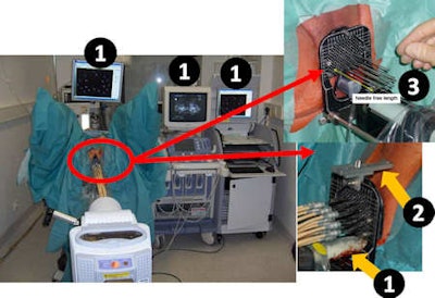
 Clinical procedure at Offenbach Clinic, showing the QA-Tools for HDR brachytherapy of prostate cancer. (1) Intraoperative real-time treatment planning system Oncentra Prostate (Nucletron) using 2D and 3D ultrasound imaging. The ultrasound probe remains in place during treatment enabling 2D and 3D verification at any time. (2) Template-Perineum QA tool. (3) Measurement of free length of the needles is utilized for both reconstruction (of needle tip) and quality control for checking needle shifts during treatment. Image courtesy of Dimos Baltas, Klinikum Offenbach.
Dimos Baltas, PhD, director of the department for medical physics and engineering of the Klinikum Offenbach's department for radiation oncology, discussed brachytherapy. He began his presentation by considering some analogies between external-beam radiotherapy and modern brachytherapy. For example: the beams are equivalent to the brachytherapy needle or catheter; beam shaping corresponds to the source stepping pattern; and raster scanning is equivalent using several catheters to cover a target volume.
The physics of dose delivery, however, differs between the two techniques. In brachytherapy, the source is so close to the target that the delivered dose is dominated by the source dwell time, and the dose distribution is more similar to the spot (Bragg peak) delivered by an ion beam. Brachytherapy is also affected by the interconnection between the source and the patient anatomy, unlike external-beam radiotherapy in which beam shaping occurs externally.
In basic terms, brachytherapy is performed by creating a pre-plan, implanting the applicator under image guidance, and then treating the patient. But while imaging patient anatomy prior to and during catheter implantation enables accurate source placement, beam's-eye-view imaging is not possible, and the catheter itself can alter the patient anatomy.
One problem arises because pretreatment imaging is usually performed in a separate room from the treatment suite. Baltas described a brachytherapy study comparing catheter positions at CT planning and during treatment. The researchers noted displacement in the few hours between the planning scan and treatment delivery and recommend verification of catheter positions immediately prior to treatment.
To do this, Klinikum Offenbach employs ultrasound imaging during brachytherapy delivery. Baltas noted that this results in a mean shift of anatomy and needles of as low as 1.0 mm. For high-dose-rate brachytherapy, this corresponds to dosimetric changes of less than 5%, implying that quality assurance measures must be implemented to achieve this one millimeter stability. It's also vital that there is no motion of the implant and anatomy prior to treatment delivery, for example, caused by removing the ultrasound probe.
Baltas rounded off his talk by considering the symposium's key question: "Can we deliver the dose distribution we plan?" Under specific conditions and scenarios, he said, data exist to support the answer "yes." However, he added, there's currently no technology available to routinely answer this for different localizations. There's also no way to measure the delivered fluence of a brachytherapy treatment; effort is needed to develop in vivo dosimetry that can perform such verification.
© IOP Publishing Limited. Republished with permission from medicalphysicsweb, a community website covering fundamental research and emerging technologies in medical imaging and radiation therapy.


