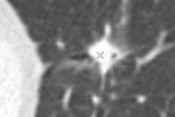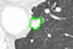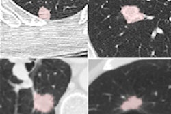
Adaptive statistical iterative reconstruction (ASIR) technology not only improves CT image quality but also increases the sensitivity of computer-aided detection (CAD) software for detecting lung nodules, according to an article published online in the European Journal of Radiology.
A research team from Osaka University Medical School in Osaka, Japan, found that applying CAD to CT data processed with ASIR reconstruction yielded significantly higher CAD sensitivity than CAD applied to conventional CT data. These sensitivity gains were found when on both standard- and low-dose CT exams.
"The sensitivity of CAD at 100%-ASIR on lower-dose CT is almost equal to that at 0%-ASIR on clinical routine-dose CT," wrote a group of authors led by Dr. Masahiro Yanagawa, PhD.
Hypothesizing that the use of MDCT with garnet detectors and ASIR might influence the ability of CAD in detecting lung nodules, the researchers sought to evaluate ASIR's effect on CAD in both standard and low-dose CT studies. They prospectively studied 35 patients (18 men and 17 women) with a mean age of 64.6 years (EJR, article in press, 5 October 2011).
Chest CT data were acquired at standard dose (120 kVp with automatically adjusted tube current) and low dose (120 kVp, 100 mA) on a Discovery CT750 HD 64-slice MDCT scanner (GE Healthcare) between June 2009 and September 2009.
The scanner's high-resolution scan mode with 2,496 views per rotation was utilized, according to the researchers. Each 0.625-mm-thick image was then reconstructed using a high-definition bone reconstruction kernel at 0%, 50%, and 100% levels of ASIR processing.
Two independent chest radiologists (with nine years and 18 years of experience, respectively) read the entire set of images without ASIR on standard-dose CT in order to create the reference standard for the study. A consensus panel, which included the same two radiologists and an adjudicator with 16 years of experience, then classified all of the candidate lesions as either true-positive or false-positive findings.
They found 265 noncalcified nodules with a diameter of ≤ 30 mm, including 103 ground-glass opacity nodules, 34 part-solid nodules, and 128 solid nodules. The nodules had a mean diameter of 4.37 mm ± 3.16 mm.
The researchers then applied CAD using the Lung VCAR software on GE's Advantage Workstation 4.2, and retrospectively assesse the CAD results in comparison with the reference standard results.
The study team found a statistically significant increase in sensitivity between the different levels of ASIR (p < 0.001).
CAD sensitivity for overall nodules
|
In particular, the researchers discovered that increasing degree of ASIR led to a significant increase in sensitivity for ground-glass opacity (GGO) nodules on standard-dose studies and solid nodules on low-dose studies.
CAD sensitivity by nodule type
|
Differences in sensitivity for GGO by ASIR type were statistically significant (p < 0.001).
On the downside, ASIR was associated with an increase in mean false-positive findings per exam, climbing from 4.6 with no ASIR on standard-dose CT studies to 8.5 with 100% ASIR, and 3.5 with no ASIR on low-dose CT exams to 6.2 with 100% ASIR. These increases also were statistically significant (p < 0.001).
Nonetheless, ASIR is a technique that can increase CAD's sensitivity for pulmonary nodules despite its increase in false-positive results, and may have the potential to improve nodule detection, even on lower-dose CT studies, the researchers concluded.
"Considering the ALARA [as low as reasonably achievable] concepts, reducing radiation dose is very important in clinical settings," they wrote. "Further analyses using various clinical data will be necessary to examine the utility of the ASIR for radiation dose reduction."
Editor's note: Thumbnail image on the home page shows PET/CT performed to stage known non-small cell lung cancer of left lung. PET/CT showed a tumor in left lower lung and subcarinal and contralateral mediastinal lymph-node metastasis (N3 stage). Image courtesy of Dr. Hans Steinert, Zurich.


