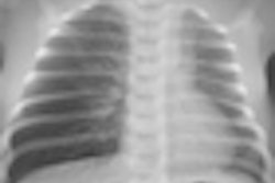
The authors of a French-language article in press published by the influential journal Archives de Pédiatrie (28 July 2011) have called for further prospective and randomized studies in children of all ages to confirm the tendency for not systematically performing an x-ray in the diagnosis of mild pediatric pneumonia.
The French National Authority for Health (HAS) advocates radiological confirmation of suspected cases, but doctors tend not to systematically perform chest x-rays in mild cases of suspected community-acquired pneumonia in children, according to lead author Dr. Rafik Bourayou, from the Service des urgences pédiatriques et de pédiatrie générale, CHU de Bicêtre, Le Kremlin-Bicêtre, France.
Chest x-ray remains a good means of etiological diagnosis, particularly in atypical presentations, and doctors are unanimous about its diagnostic role in severe infection. In particular, chest x-ray should be performed if a child has signs of pleurisy during a clinical examination, nonimprovement of symptoms after 48 hours, significant weight loss, hemoptysis, or underlying chronic illness, among many other factors, he noted.
The benefits of using chest x-ray in diagnosing mild pneumonia, however, remain unclear, and opinions vary about its exact role in current practice.
Based on South African research on the outcomes of children up to age 59 months with acute lower respiratory infection, in 2002 the British Thoracic Society published recommendations to not systematically x-ray nonhospitalized children with mild infection. The move was quickly followed by bodies in Canada and the U.S. However, in 2009, HAS declared that chest x-rays should be performed in children with suspected pneumonia, as there was otherwise insufficient proof to exclude it. On the other hand, HAS does not recommend follow-up chest x-rays, particularly if the patient seems to be clinically improving.
It is widely accepted that prolonged or frequent ionizing radiation from x-rays leads to an accumulative risk of cancer. The damaging effect of a few low-dose studies has not been determined, but constitutes one reason for curbing examinations, according to Bourayou.
Cost is another issue. In France, a chest x-ray costs between 21 and 40 euros ($29 to $56 U.S.). Staff at CHU de Bicêtre's emergency pediatric service perform between two and 12 a day to diagnose pulmonary origin infection, resulting in an annual expenditure of about 80,000 euros ($113,000 U.S.) for this indication.
To reduce the number of chest x-rays carried out for diagnosing pneumonia, research has sought to determine how clinical assessment of criteria, such as lowered vesicular murmur, can exclude the infection, but such methods to date suffer from a lack of sensitivity.
In suspicious cases, attempts to better define factors predictive of the radiological presence of infiltrates for a more tailored approach to patient selection have met with limited success. While these criteria can be identified and measured to yield high predictive sensitivity, specificity remains poor. Nor can chest x-ray reveal causal agents of infection -- viral, bacterial, or atypical -- and so cannot guide therapy, the authors wrote.


