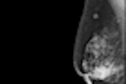
Poor mammography image quality was found at several centers in a prospective, 17-country study by Serbian researchers published in the European Journal of Radiology. However, simple corrective actions such as cleaning screens can result in large improvements, they found.
As many countries move toward a population-based breast screening program, it is important to ensure good image quality so patients are not exposed to unnecessary radiation, the authors noted. The European Council Directive 97/43/Euratom and the International Atomic Energy Agency's (IAEA) International Basic Safety Standards for Protection Against Ionizing Radiation and for the Safety of Radiation Sources require standardization of imaging techniques and other procedures involved in mammography.
The IAEA initiated a project to assess the mammography situation in various countries as part of its program on radiation protection of patients. The project sought to evaluate optimization of protection in clinical mammography examinations, identify areas where action is needed, and then document improvements after corrective actions are put in place. Dr. Olivera Ciraj-Bjelac from the Vinca Institute of Nuclear Sciences in Belgrade, Serbia, and colleagues surveyed 17 countries for their study: Ghana, Tanzania, Uganda, Iran, Malaysia, Pakistan, Syria, Bosnia and Herzegovina, Bulgaria, Croatia, Greece, Lithuania, Macedonia, Malta, Moldova, Romania, and Serbia (Eur J Radiol,12 June 2011).
In the first phase of the prospective trial, the researchers assessed the initial image quality of clinical images and identified the causes for poor image quality during a two-week period. One radiologist and one radiographer were assigned to determine the image quality for each mammography unit.
For 54 mammography units, the total number of images graded in the first phase of the project for craniocaudal and mediolateral oblique projection was 6,572 and 6,492, respectively. For the craniocaudal projection in this phase, the fraction of images assessed without any remark (A grade) was 70% (average) with a range of 13% to 100%. The percentage of images with at least one remark (B and C grades combined) was 30% on average, with a maximum of up to 86%.
For the mediolateral oblique projection in this phase, the fraction of images assessed as A grade was 72% (average) with a range of 2% to 100%. The percentage of images with at least one remark (B and C grades combined) was 28% on average, with a range from negligible to 82%.
The main causes for poor image quality were artifact, overexposure, or underexposure for both projections. Based on the results of the initial image quality assessment, corrective actions were proposed and explained to the staff by a local medical physicist. After implementation of corrective actions, image quality was reassessed in the same mammography units and using the same methodology for another two weeks, as a second part of the study.
Only 24 units from 10 countries (eight from Europe, one from Africa, and one from Asia) participated in the second phase of the study, which included implementation of corrective actions and reassessment of image quality, as well physical evaluation of image quality and dose. Types of corrective action were based on the problems that caused the degradation of image quality. Major problems were related to film processing, damaged or scratched image receptors, film-screen combinations that are not spectrally matched, inappropriate radiographic techniques, and lack of training.
For the 24 mammography units, the total number of images graded in the second phase of the project for craniocaudal and mediolateral oblique projection was 3,845 and 4,420, respectively. After implementation of corrective actions, the percentage of A grade images for craniocaudal projections increased by nine points to 80%, while the percentage of B and C grades dropped to 20%.
For mediolateral oblique projections, after implementation of corrective actions, the percentage of A grade images rose by seven points to 83%, while the percentage of B and C grades dropped to 17%.
The researchers identified the following simple but effective corrective actions (mainly related to technique and the operator):
- Cleaning of screens, cassettes, processing units, and working surfaces (in eight units)
- Improvement of exposure technique by using automatic exposure control (aec) instead of manual selection of exposure parameters
- Improved compression and tube voltage settings (in 21 units)
- Introduction of quality control activities such as sensitometry (in six units)
- Replacement of cassettes and image reception systems (four units)
- Improvement of viewing conditions (in three units)
Other corrective measures included introduction of quality control on a daily basis, improvement of image viewing conditions and compression devices, and training of radiographers.
"Although these actions do not require large investments, they can equally or more contribute to improved image quality compared to actions that require large investments (replacements of image receptors, processing units, or even mammography x-ray units)," Ciraj-Bjelac and colleagues wrote. "Simple, low-cost, and immediately implemented measures resulted in more significant improvements of image quality compared to more costly measures. In some centers, an improved image quality grading by 50 percentage points was observed, when low-cost corrective measures were applied."
The results of the study confirm a need to evaluate the current situation concerning optimization of protection in mammography in different countries, identify the areas where action is needed, and document improvement after corrective actions are put in place, the authors noted.
"An increased awareness of the importance of producing good-quality mammograms and, thus, preventing unnecessary patient exposure should be a goal in all countries," they wrote. "This is of particular importance for countries that are moving toward introduction of population-based screening programs in mammography."


