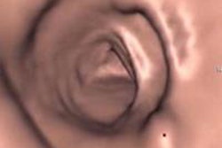
Size and morphology are good predictors of whether a colorectal lesion harbors advanced histology and needs to be removed, according to a new multicenter study from Italy. The results suggest that polyps of the same size detected at virtual colonoscopy could be classified and managed differently based on their morphology.
"In the literature there is ongoing debate about what we should do with polyps ranging from 6 to 9 mm. Should we send them for immediate polypectomy, or should we adopt a more conservative approach?" said Lia Morra, Ph.D., from computer-aided detection (CAD) firm im3D of Turin, Italy.
In a presentation at the 2009 RSNA meeting, Morra discussed the group's work in trying to understand whether using VC (also known as CT colonography or CTC) imaging features other than polyp size, such as morphological features of the polyp, could help them recognize which polyps should be sent to immediate polypectomy.
The group's larger goal was to develop more effective strategies for managing small polyps detected at CTC by developing a model to predict advanced neoplasia versus other histological types based solely on CTC characteristics.
IMPACT data
From data gathered throughout Italy in the multicenter IMPACT screening trial, the group enrolled more than 700 high-risk patients, of which more than 200 had a polyp detected at conventional or virtual colonoscopy, she said. Data analyzed for the study included 235 polyps ≥ 6 mm that had been confirmed at conventional colonoscopy from 170 subjects in the trial.
Two experienced radiologists reviewed the cases with all available information including size, location, and morphology of the lesions. Each polyp was defined as advanced neoplasia, which included adenomas and cancers, and subdivided further into low-risk adenomas and hyperplastic polyps. Flat polyps were defined as those three or more times longer than their height.
Using CTC characteristics alone, the researchers developed a logistic regression model to discriminate lesions smaller than 3 cm at CTC as advanced neoplasia (AN) or other histotypes (OH). The data were randomly divided into training sets based on 150 randomly extracted lesions (89 AN, 61 OH) and testing (n = 89) datasets for development of the model. Finally, the feature predictiveness was examined using a chi-square test. Model accuracy was verified using a receiver operator characteristics (ROC) curve on the remaining 85 lesions (53 AN, 32 OH).
Next the researchers applied the model to classify the data into the AN/OH groups based on their morphology (pedunculated, sessile, or flat), defined as base greater than twice the height and size, measured as the largest lesion diameter at the prone or supine 2D view. Based on the CTC features, the model predicts the probability of each lesion being an AN.
According to histological results, 142 lesions were AN (advanced adenoma/cancer) and the remaining 93 were OH (low risk/hyperplastic polyps).
"If we analyze the distribution of both size and shape, not surprisingly, most larger polyps are actually advanced neoplasia," Morra said. "However, also one-third of medium-sized polyps are also advanced neoplasia, and we did not want to miss them. Both CTC size and morphology were found to be significant predictors of advanced neoplasia [p < 0.001], and I think CTC with morphology as a model actually improves the detection of advanced neoplasia."
Pedunculated polyps
Morra said that specifically, the pedunculated polyps actually had a higher probability of being advanced neoplasia than other types (p < 0.001), and this is especially true in the medium-sized polyps.
The model's estimated probability of a medium-sized (6-9 mm) pedunculated polyp having advanced morphology is higher than that of sessile or flat lesions (p = 0.003). For example, an 8-mm pedunculated polyp has a probability of harboring advanced neoplasia of 74% (± 9% standard deviation [SD]), versus 36% (± 6% SD) and 22% (± 10% SD) for sessile and flat polyps of the same size, respectively.
CTC size and morphology were significant predictors of AN (p < 0.001); however, adding morphological features to the analysis significantly improved its predictive capacity (p = 0.002), the researchers wrote in an abstract.
The ROC area under the curve was 0.84. At an operating point that corresponded to a good compromise between sensitivity and specificity, 47/53 advanced neoplasia (89%, confidence interval [CI]: 77%-96%) and 21/32 lesions of other histotypes (65%, CI: 47%-81%) were classified correctly for an overall diagnostic accuracy of 81% (CI: 70%-88%).
"We can probably be more aggressive with [removing] pedunculated polyps and less aggressive with the sessile polyps," Morra said. "The results suggest we should use a different size threshold for referral for polypectomy depending on polyp morphology."
Without the model, referring all polyps 10 mm or larger for immediate colonoscopy would yield a sensitivity of 64%, "which is actually quite lower than the 81% that we obtained here," Morra said. Specificity was marginally higher using the model, 89% versus 75%."
"We have shown that CTC morphology can be used along with size to allow better discrimination of advanced neoplasia than when using polyp size alone," Morra said.
Morra went on to state that researchers need a model where polyp-specific information is complemented by patient-specific information to discriminate between high-risk and low-risk patients, and actually identify the people most likely to benefit from polypectomy.
After the talk, Dr. Abraham Dachman from the University of Chicago commented on the difficulty of classifying polyps precisely into morphologic categories.
"Looking at datasets from multiple institutions sent to me for other purposes, one of the things I noticed was the different way people use the word 'pedunculated,' " Dachman said. "We know about the controversy over the definition of a flat lesion, but I also find it interesting that when something is truly spherical, everybody says sessile, but when it starts to get a little bit of a neck, some people call it sessile and some call it pedunculated."
Morra responded that the group did not obtain detailed information on the criteria the researchers used to classify the polyps.
By Eric Barnes
AuntMinnie.com staff writer
December 22, 2009
Related Reading
CAD catches most flat polyps on virtual colonoscopy, December 16, 2009
ACRIN: Virtual colonoscopy sensitive for flat polyps, October 13, 2009
Flat-polyp measurements show less variability in 3D VC, September 16, 2009
Virtual colonoscopy CAD improves flat-lesion detection, June 5, 2009
ACRIN trial shows VC accuracy comparable to optical colonoscopy, September 18, 2008
Copyright © 2009 AuntMinnie.com


