Second-look breast ultrasound with volume navigation effectively detects occult additional lesions initially spotted via breast MRI, Italian researchers have found. In addition, the technique permits safe biopsies irrespective of position and depth.
In a study of nearly 2,000 breast MRI exams, Dr. Alfonso Fausto from the department of diagnostic imaging at Azienda Ospedaliera Universitaria Senese in Siena, Italy, and colleagues prospectively evaluated second-look ultrasound using volume navigation for MRI-detected additional breast lesions. Second-look ultrasound with volume navigation identified 151 additional lesions, they found.
"This could be a reliable technique in view of the technological improvement yielding high image quality of breast MRI in supine position," they wrote (European Radiology, 15 October 2018). "The application of this technique makes it possible to detect a large number of additional breast lesions, occult at second-look ultrasound, and to biopsy a significant number of malignant lesions safely and irrespective of the distance from the skin or lesion position."
Take a second look
Second-look breast ultrasound is often used in clinical practice to reassess a patient after detecting additional lesions via another technique, such as contrast-enhanced MRI. Targeted second-look ultrasound makes it possible to identify correlated lesions at a pooled rate of 57.5%, according to Fausto and colleagues.
A new technique that combines live ultrasound of the breast and MR imaging obtained in supine position with similar parameters to the prone examination could help radiologists with their endeavor to identify and treat breast cancer.
"A new world has suddenly opened up where the intrinsic advantages of the two methods, ultrasound and MRI, are combined with the aim to see and perform what until then was impossible: an objective second-look ultrasound of enhancing MRI-detected lesions using MRI volume as a 'road map,' and, when necessary, perform a biopsy during an ultrasound exam with MRI guidance. A real revolution!" Fausto told AuntMinnieEurope.com.
He and his colleagues codified the way to coregister live ultrasound exams with previously acquired breast MRI volume, which they're calling breast ultrasound with MRI volume navigation.
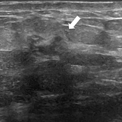
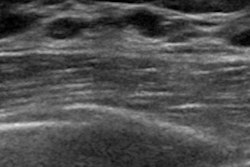
 Ultrasound image (left) with the corresponding multiplanar reconstructed MR image (right) of a 54-year-old woman with a suspicious lesion at the upper-internal quadrant of the right breast. MR imaging showed an irregular branched enhancing lesion of 23 mm, undetected at second-look ultrasound. Second-look ultrasound with volume navigation showed the corresponding area (white arrow). Pathology obtained with ultrasound-guided biopsy with volume navigation demonstrated an invasive ductal carcinoma with an extensive intraductal component (green box). All images courtesy of Dr. Alfonso Fausto.
In the current study, they present the results of a six-year assessment of second-look ultrasound of additional breast lesions with volume navigation using supine contrast-enhanced MRI coregistered during live ultrasound exams to analyze its clinical value using pathology as a reference, and to assess the need for MRI-guided biopsies.
The study took place from January 2009 to December 2015 and includes 1,981 1.5-tesla MRI exams (Achieva, Philips Healthcare; Signa HDxt, GE Healthcare) in 1,437 patients. Before each exam, all patients underwent a clinical exam and conventional imaging with mammography and ultrasound.
For patients with an additional lesion, a second-look ultrasound was performed by the same radiologist who evaluated the MRI examination. Ultrasound-guided biopsy was immediately performed in the case of concordance between second-look ultrasound and MRI.
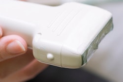
Live ultrasound exams were performed using a platform configured with volume navigation (Logiq E9, GE Healthcare) and using a 6- to 15-MHz transducer with a geometry permitting visualization of a wide field-of-view. Both ultrasound and MR images were displayed side by side or in a blended overlaying format on the ultrasound monitor. After coregistration, an ultrasound-guided biopsy with volume navigation was immediately performed systematically leaving a tissue marker of pyrolytic carbon-coated zirconium oxide before ending the procedure.
Fausto and colleagues found in 490 MRI examinations that at least one additional breast lesion was detected, for a total of 722 only MRI-detected lesions. Results are summarized in the table below.
| Additional lesions identified & types | |||
| Ultrasound-guided biopsy | Ultrasound-guided biopsy with volume navigation | MR-guided biopsy | |
| Benign | 362 | 67 | 20 |
| Malignant at pathology | 187 | 84 | 2 |
An additional lesion after breast MRI is not rare -- in this study, it occurred in about 25% of all MRI exams. For these lesions, a second-look ultrasound is mandatory. However, due to the large variability of displacement of the breast tissue, the chance of finding a lesion correlation during a second-look ultrasound exam ranges from 58% to 64%, according to the researchers.
"An MRI-guided procedure is undoubtedly the primary technique used to obtain pathology results from a lesion occult at second-look ultrasound," they wrote. "However, the cost of the examination in terms of the time for which the MR equipment is used (from the performance of the biopsy up to reconditioning of the premises), and of MRI-compatible materials is certainly higher, especially in relation to the high rate of false positives."
Also, the inability to reach lesions located near the chest wall or the nipple area, and the high degree of expertise required to perform it, make the development of a complementary method necessary, they added. The current research demonstrates ultrasound-guided biopsy with volume navigation could be that method.
"In our study, only 22 lesions underwent MRI-guided biopsy in six years, demonstrating the reduced need for this technique," the study authors wrote. "Moreover, in view of the scarcity of centers performing MRI-guided biopsies, this new technique could assume an important role, offering an alternative to MRI-guided biopsy for a large number of additional lesions."
Future research will focus on patient position during breast MRI: If the supine position can demonstrate the same diagnostic value as the examination in the prone position, an "objective" second-look ultrasound or a similar approach could easily be performed also in the operating room.
"If our hypothesis is correct, it would certainly be easier to coregister MR images during the ultrasound examination, without requiring any additional imaging," Fausto told AuntMinnie.com. "But it is not enough. The benefits could be larger showing breast lesion localization during the surgical intervention, quantifying tumor detection of residual breast cancer, and, finally, to guiding radiotherapy treatment in patients undergoing conservative surgery."


