Dear Cardiac Imaging Insider,
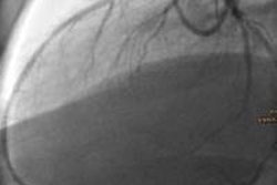
In recent years, a broad range of techniques have been developed to determine whether a coronary artery stenosis detected at CT angiography is hemodynamically relevant -- or whether it only looks that way and doesn't affect blood flow.
The techniques include algorithms that calculate fractional flow reserve based on CT images, with one such protocol requiring CT data to be sent for remote computation. Then there is the simpler method of quantifying the lumen area based on reconstructed CT images of stenosed coronaries, which hadn't been tried before. Dr. Fabian Plank and colleagues from Innsbruck, Austria, honed such a technique into a fast, highly sensitive, and specific way to identify flow-limiting lesions. Click here for more.
MRI is the modality of choice for imaging cardiac veins, congenital heart disease, and masses, but CT's star continues to rise in assessment of the coronary arteries, according to a presentation at this week's Arab Health Congress in Dubai, United Arab Emirates. As the machines that acquire the images have evolved, cardiac CT has also been shown to reduce the costs of care in several important ways. For the rest of the story, click here.
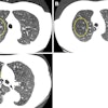
Coronary calcium scores are a simple and highly validated way to assess the risk of heart attack. But scanners are all over the map in reporting them, according to Dutch researchers who keep finding differences in how CT measures coronary calcium. In their latest study, they put four scanners to the test on a bit of calcium in a phantom. Not only were the scores widely divergent, but patient size mattered a lot.
A little motion also throws calcium scores into havoc, according to Dutch researchers on a mission. The effect of motion on calcium is so large it even dwarfs the substantial effects of iterative reconstruction, they report here.
The U.K. has published new guidelines on the safe use of coronary CTA. Get the details here.
Researchers in France have developed an ultrasound method that can produce 3D videos at thousands of frames per second, enabling all kinds of new tricks such as watching shear waves flowing through the heart. Check out ultrafast 3D ultrasound here.
Finally, we invite you to scroll through the links below for the rest of the news in your Cardiac Imaging Community.


